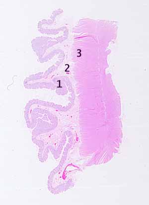
The source for this specimen should be obvious (and confidently so), provided you are familiar with the organ it represents.You should easily see three distinct layers, 1 mucosa, 2 submucosa, and 3 two-layered muscularis. Only one organ system has such distinct layers.
The mucosa is unique. Note that the mucosa contains numerous tubular invaginations, which extend from the surface all the way to the muscularis mucosae. Also note that lamina propria is conspicuous between these invaginations.
Note that one type of columnar epithelium lines both the surface and the invaginations. Many of these epithelial cells contain a large, unstained "bubble" (maybe a mucus droplet?).
Only one organ has features like this.
No more hints.
