This slide presents some peculiar difficulties, but also abundant
clues for correct identification.
Follow the dotted line at right to see how a rather thin tissue specimen has been folded.
This leads to a somewhat confusing appearance, especially in the region
indicated by number 1.
However, if you use your imagination to "unfold" the tissue,
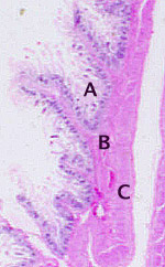
you should easily see three distinct layers, labelled
A,
B, and
C in the enlargement
of region 2.
- A mucosa
- B submucosa
- C two-layered muscularis
Only one organ system has such distinct layers.
 The enlargement of region 2
illustrates several folds of mucosa.
Each such fold (one example extends leftward and upward from letter
B) has a core of submucosal connective tissue that is more substantial than lamina
propria. Such folds can be found all along the mucosal surface.
Most of the surface epithelium has been lost, leaving behind a "naked" lamina propria.
(This is a fairly typical post-mortem change.) The shape of the remaining rather tenuous
wisps of lamina propria reveals
the original shape of the mucosa, especially if you use your mind's eye to cover
each remaining shred of lamina propria with epithelium. (Some characteristic epithelium can still be easily
found in protected sites deep in the mucosa.)
Use the three distinct layers, the appearance of the remaining epithelium,
and the charactistic shape of the lamina propria and of the
submucosa to deduce which region of which organ system this must be.
No more hints.
