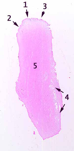
You should be able to see a stratified squamous epithelium in the region labelled with number 2.
The stratified squamous epithelium ends at number 1.
Beyond point 1, in the region labelled 3, you may be able to find scattered columnar cells.
Although most of the columnar epithelium has been lost (a fairly typical post-mortem change), you can nevertheless reliably notice the end of the stratified squamous epithelium. Furthermore, you can also reliably notice a marked transition in the underlying lamina propria, with many more lymphocytes appearing under the columar epithelium than under the squamous epithelium.
So, this specimen comes from some location where a stratified squamous epithelium gives way to a more delicate epithelium. List all such locations. (Basically, consider every orifice of the body.)
Which of these locations is/are consistent with the smooth muscle that is visible throughout the bulk of this specimen (number 5), with the extensive vasculature in the lamina propria under number 2 and with the glands lined by columnar mucous cells at number 4?
No more hints.
