The source for this specimen should be obvious (and confidently
so), provided you are familiar with the organ it represents.
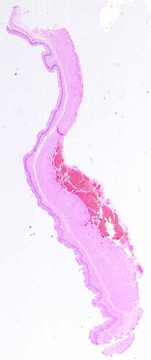
Note the various distinct layers.
Which organs have such distinct layers?
Note specific features of the mucosal layer.
- What shape is the surface?
- How many different kinds of epithelial cells comprise the surface
epithelium?
- Make sure you examine the entire epithelial surface.
- Are there any epithelial features (e.g., glands or crypts) embedded
in the lamina propria?
- Can you see a distinct muscularis mucosae?
Are there any distinctive features in the submucosa?
Is the muscularis externa organized into distinct layers? What
kind of muscle comprises the muscularis in this specimen?
What is the bright red stuff deep to the muscularis?
This slide should be easy. The type of surface
epithelium, together with the appearance of distinct layers -- mucosa
with thick muscularis mucosae, submucosa, and muscularis externa --
is characteristic of only region of the body. (The bright red
stuff is spilled blood.)
There are no more hints.