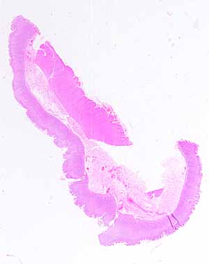
The source for this specimen should be obvious (and confidently so), provided you are familiar with the organ it represents.Note the several distinct layers.
- Name these layers.
Which organs have such distinct layers?
Note specific features of the mucosal layer.
- What shape is the surface?
- How many different kinds of epithelial cells comprise the surface epithelium?
- Is lamina propria conspicuous?
- Are there any epithelial features (e.g., glands or crypts) embedded in the lamina propria?
- Can you find a muscularis mucosae?
Are there any distinctive features in the submucosa?
Is the muscularis externa organized into distinct layers?
Hints on the next page are a bit more pointed. Don't look unless you are stuck.
