The source for this specimen should be obvious (and confidently
so), provided you are familiar with the organ it represents.
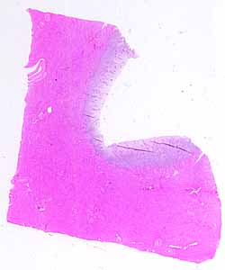
Note the cut and uncut surfaces of this specimen.
Note the quality of tissue near the uncut surface.
- What kind of epithelium is present?
- Observe the glandular structures. Note their shape and their
epithelial lining.
- Note the amount and quality of interstitial (stromal) tissue. Does
this look like lamina propria?
Note the thickness and the texture of the deeper tissues (beneath the
epithelium, glands, and associated stroma).
- What kind of tissue comprises the bulk of this deeper layer?
- How is this tissue organized (i.e., in layers or in variously interwoven
bundles)?.
Which organ has features such as these?
Do the epithelial cells appear actively secretory? Can you find
mitotic figures in the glandular epithelium? What state of activity
is suggested by these details?
No more hints.