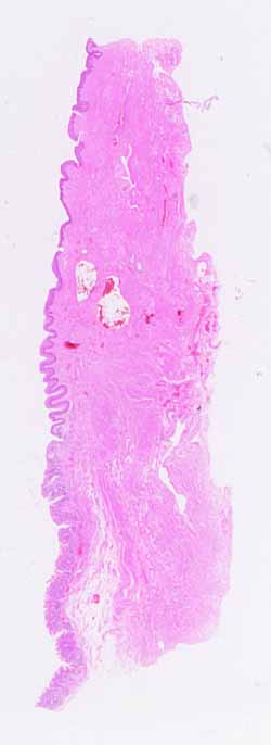
The source for this specimen should be obvious (and confidently so), provided you have noticed all the conspicuous clues.Hopefully, you have found stratified squamous epithelium at one end of the specimen and columnar epithelium at the other end.
- The transition from stratified squamous to simple columnar is abrupt. Unfortunately, on this specimen the surface columnar cells are missing at the site. (This is a common post-mortem change.)
- There are only a few locations where such a transition occurs. This specimen must come from one of them.
Less obviously (but we hope you've already noticed), the stratified squamous epithelium differs between the cut end of the specimen and the transition to columnar. If you haven't already found this difference, do so now.
- The transition from non-keratinized to keratinized epithelium is gradual but nevertheless easy to recognize. Look for the presence or absence of a stratum granulosum as well as a stratum corneum. (If these latter terms are unfamiliar, review epidermis of skin.)
- There is only one place in the body where epithelium shows all of these changes -- from simple columnar to non-keratinized to keratinized -- over such a short distance.
The difference in underlying tissue layers, both connective tissue and muscle, correlates nicely with the epithelial transitions.
- From the end of the specimen where a distinct muscularis mucosae appears, follow the muscularis mucosae and notice where it stops (relative to changes in the overlying epithelium).
- Similarly, notice where loose connective of submucosa gives way to more densely fibrous connective tissue.
- Finally, notice how a distinct muscularis layer becomes less distinct and more interwoven with the densely fibrous connective tissue.
No more hints.
