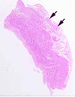
This may be one of the more difficult specimens in the SAQ series. There is, however, one small region that offers a reliable clue for locating the source.Look at the site indicated by the arrows, in the adventitial layer. Can you find conspicuous tubular structures?
For these tubular structures, note the epithelium, the shape of the lumen, and the thickness of the muscular wall relative to the lumen. Only one particular part of the body has this set of features. If you have studied that part, these tubular structures should be recognizable (they look just like the textbook pictures, and the reference slide).
In this specimen, these small tubes are attached to a much larger organ, one with a muscular wall and a mucosal surface. So, now you only need to figure out where such tubes run alongside an organ with such a wall. And make sure that all the various details are consistent.
[Incidently, at least some slides from this specimen include a parasympathetic ganglion. Look between the tubules and the wall of the adjacent organ.]
No more hints.
