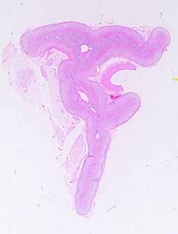
This should be the easiest specimen of the SAQ series. It can be confidently and reliably identified WITHOUT use of microscope, based on shape alone. But use your microscope anyway.Note the connective tissue capsule with many associated blood vessels and nerves around the periphery of this specimen.
As you scan across this specimen from one side to the other, note that the texture of the parenchymal tissue of the outer portions is distinctly different from that of the inner portion. This difference between outer and inner portions is most obvious in the thickest region of the specimen, in the vicinity of the larger internal blood vessels.
Note that the tissue comprising the outer portion may be divided into indistinct zones, based on parenchymal cell arrangement.
Note the peculiar bands of longitudinal muscle (cut in cross section) in the walls of the veins in the inner region.
[Incidently, look for unusual texture in the adipose tissue around this organ. This includes not only ordinary white fat but also brown fat (multilocular fat), used for heat generation.]
No more hints.
