This specimen should be readily recognizable, provided you
are familiar with the organ it represents.
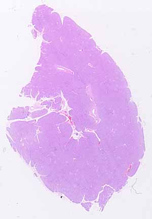
Note that this is a solid organ (as opposed to a hollow or tubular
one).
Note that the parenchymal tissue is organized into lobules of various
sizes, separated by thin strands of connective tissue.
Note the many small tubular structures scattered throughout the specimen,
with larger tubules in the thicker masses of connective tissue.
- What kinds of epithelium line these various tubules?
Note that the parenchyma comprising the lobules consists of very many
small clusters of cells. In each cluster, the inner end of each
cell appears eosinophilic and grainy while the outer end appears basophilic.
Hints on the next page are more revealing. Don't look until
you are ready to confirm your identification.