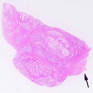
Hopefully, you have identified this specimen as glandular tissue consisting of many irregular tubules with a convoluted epithelial lining, within a relatively thick stroma.
Note the appearance of the secretory epithelium, consisting of columnar cells with fairly clear apical cytoplasm. Also note the occasional appearance of acidophilic concretions within the lumens of some secretory tubules.
The stromal composition is significant, but somewhat difficult to recognize with confidence. (A trichrome stain would have helped. Sorry.) Note that some of the fibers in the stroma are actually cells (rather than extracellular collagen), with nuclei within the eosinophilic fibers.
Only one large gland has such a texture of irregular tubules lined by convoluted, columnar epithelium, within a fibromuscular stroma.
All of the boundaries of this specimen show cut surfaces, but the connective tissue capsule of the organ, with a few striated muscle fibers in the adventitia, may be seen in the region indicated by the arrow. The striated muscle assures that this specimen did not come from within the peritoneal cavity.
No more hints.
