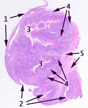
The source for this specimen should be obvious (and confidently so), provided you are familiar with the organ it represents. The sample is normal, not pathological.
- In what parts of the body does a nonkeratinized stratified squamous epithelium occur? (arrow 1)
- Where does such a stratified squamous epithelium turn inward to form conspicuous crevices? (arrow 2)
- Note that within the deep crevices (2 and 3) the epithelium becomes obscured by infiltration with many small cells. What are these small cells, and what are they doing here?
- Note the extremely numerous small cells with small round nuclei throughout the solid portion of the specimen. Observe that these cells are organized into prominent pale patches (arrow 4) with darker, more-densely-packed caps. (Again, what are these small cells, and what are they doing?)
- Finally, note that the deep, supporting tissue includes not only fibrous connective tissue but also striated muscle (arrow 5).
This slide should be easy. The presence of so many lymph nodules in a site covered by stratified squamous epithelium with deep crevices is characteristic of only one region. Other details of infiltrating cells and deeper tissues are consistent with this site.
No more hints.
