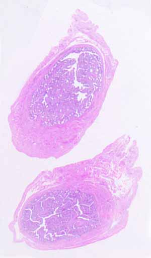
This is NOT small intestine. (It's much too small, among other differences.)
This is NOT gall bladder, either. (The epithelium is wrong, among other differences).
The most distinctive feature of this tubular organ is the presence of elaborate mucosal folds that almost completely fill the lumen.
Note that none of the mucosal folds appear detached (i.e., as free-floating "islands"); similarly, the entire complex lumenal space is interconnected (no "chambers" separate from the principal lumen). Each of these observations implies that all of the mucosal folds are longitudinal, such that all of the elaborate crevices between the mucosal folds must extend as straight passageways along the length of the organ. Thus there are no "pockets" where a travelling object might get caught.
The muscular wall is quite thin. The deeper tissues contain conspicuous blood vessels.
The epithelium consists of more than one cell type, although these cell types cannot be easily distinguished on this specimen
- Some epithelial cells have relatively large, oval, euchromatic nuclei.
- Others have relatively narrower, darker nuclei.
- Some epithelial cells (those with the larger, paler nuclei) display an apical specialization which projects into the lumen. This is NOT a brush border of microvilli. (Observe, at high power, that these apical projections are much longer and coarser than microvilli.)
