
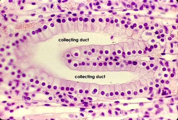
RENAL IMAGE INDEXTwo collecting ducts are merging here into a larger duct, deep in the medulla. Urine flow is toward the left. The surrounding tissue consists of medullary blood vessels and thin segments of loops of Henle. Because both are lined by simple squamous epithelium, vessels and loops can be difficult to distinguish, except when blood is present in the vessels.
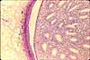
Click on thumbnails at left for lower-magnification views of this region.
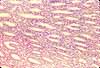
Click on the thumbnail at right for comparison of medullary tubules cut transversely.
Comments and questions: dgking@siu.edu
SIUC / School
of Medicine / Anatomy / David
King
https://histology.siu.edu/crr/RN009b.htm
Last updated: 30 May 2022 / dgk