
 Histology Study Guide
Kidney and Urinary Tract
Kidneys excrete urine. They do this by producing a modified filtrate of blood plasma.
Note that renal physiology and pathology cannot be properly understood without appreciating some underlying histological detail.
The histological composition of kidney is essentially that of a
gland
with highly modified secretory units and highly specialized ducts.
[For an overview of glandular organization, see
glands in the introductory
unit.]
Please note that this is an ancillary resource,
NOT a substitute for textbooks or for time spent studying real specimens with
a microscope. If you use
this on-line study aid, please refer to your textbooks
and atlases and resource sessions for richer, more detailed information.
Click here or scroll down to continue this introduction
to kidney. To go directly to specific topics, see the table below.
|
|
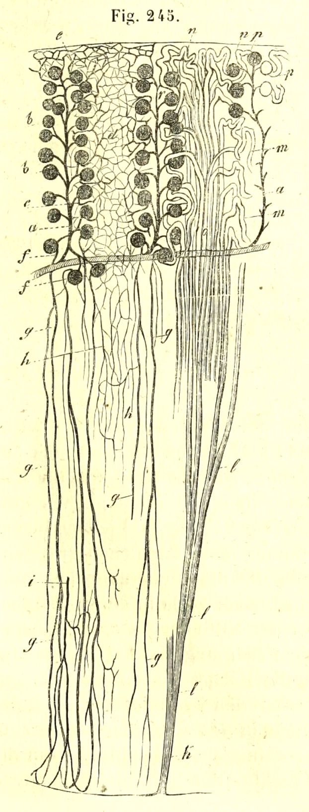 |
| Essential microanatomy of kidney, from Kölliker's
1852 Handbuch der Gewebelehre.
Vasculature is shown on left side, epithelial tubules on right. |
|
TOP OF THIS PAGE / CONTENTS
FOR THIS PAGE / RENAL IMAGE INDEX
Renal pathology
CLINICAL NOTE: From a clinical perspective, the most significant
histological feature of the kidney is probably the renal
corpuscle. Several pathological processes interfere with glomerular
filtration and thus have a critical impact on kidney function. Diagnosis
of renal pathology often involves biopsy of renal cortex and careful examination
of renal corpuscles. Corpuscle details such glomerular basement membranes, podocytes,
and mesangial cells can be revealed by several special stains
as well as by electron microscopy.
 The thumbnail to right links to some images of membranoproliferative
glomerulonephritis.
External links to WebPath
provide additional information and images:
- WebPath
electron micrographs of renal pathology
- "Minimal
change disease (MCD) with effacement of podocyte foot processes."
Note that podocytes form a continuous surface over the capillary; filtration
slits are missing.
- "Membranous
glomerulonephritis with thickened glomerular basement membrane containing
electron dense deposits."
- WebPath tutorial on renal
cystic disease.
- WebPath tutorial on urinalysis
(including brief descriptions of how urinary casts are related to glomerular
and tubular function)
- Additional renal pathology at WebPath. (This page is worth an extended visit, after you have become
comfortable with basic kidney histology.)
TOP OF THIS PAGE / CONTENTS
FOR THIS PAGE / RENAL IMAGE INDEX
Overview of kidney histology
The underlying tissue organization of the kidney is that of a compound exocrine gland
such as pancreas or salivary gland, albeit with highly modified secretory units and highly specialized ducts.
(If you are not yet familiar with glands, you might helpfully review basic patterns of
tissue organization. You needn't master all the details of glands before
you study kidney, but this page might offer a helpful sense of how epithelial cells
arrange themselves to form the kidney's essential structures of the kidney.)
Image contrast, gland vs. kidney
(click on any thumbnail for enlarged comparison)
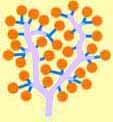 |
Gland (alveoli and ducts)
The bulk of a typical exocrine gland,
such as a salivary gland, consists
of secretory epithelial cells. These cells form secretory units,
called acini, which drain their
secretory product into a branching tree of ducts.
In this cartoon, orange color
represents acini, blue color indicates
the short striated ducts,
where secretory product is concentrated by active reabsorption, and
lavender color
indicates interlobular and interlobar ducts, whose function is more
passive.
|
|
 |
Kidney (corpuscles and tubules)
In contrast, the bulk of the kidney consists of tubules. The secretory units of the kidney, called renal
corpuscles, comprise a relatively small proportion of the organ.
Most of the kidney consists of highly specialized renal tubules,
which correspond to the duct tree of a typical gland. Together,
one renal corpuscle and its associated tubule is called a nephron.
In this cartoon, orange
color represents renal corpuscles,
blue color indicates tubules
associated with each corpuscle, whose function is analogous to that
of striated ducts,
and lavender color
indicates collecting ducts, whose function is more passive.
|
|
 |
Glandular alveolus
In a typical exocrine gland, each acinus
uses raw materials supplied by blood to manufacture a secretory product.
As this product drains away, it may be concentrated by
a striated duct.
|
|
 |
Kidney corpuscle
In the kidney, each renal
corpuscle is essentially just a highly modified secretory acinus.
However, each corpuscle "secretes" a filtrate of blood plasma which drains
into its associated renal tubule. |
|
 |
Kidney tubules
Renal tubules have wiggly cortical portions, called convoluted tubules,
and straight medullary portions, called loops of Henle and
collecting ducts. These different segments carry out different aspects of filtrate
modification. The cortical tubules, analogous to
striated ducts, modify the filtrate
by reabsorbing everything that is not waste. |
|
| | Everything in the paragraphs above is described in greater detail further down this page.
| |
|
Housecleaning
An analogy for liver and kidney function.
Although fundamentally built like a gland, the kidney functions primarily as a filter for blood.
The body contains two "blood-filter" organs, the liver and the kidney.
Both organs serve to remove unwanted materials from blood, but they do so by two very different methods. Without going
into any molecular detail, we might compare these two methods to the strategies adopted by two different householders who set out
to eliminate clutter from their respective homes.
One householder identifies each unwanted item and tosses it into the trash. Only materials identifiable as trash are disposed of.
This householder works like the liver, where hepatocytes take up various molecules
from the blood and destroy or detoxify them as needed.
The other householder first puts everything out into the yard, then identifies and retrieves anything that was worth keeping.
Only materials identifiable as valuable are retrieved. This householder works like the kidney, which lets practically everything
pass out from blood into glomerular filtrate and then uses proximal tubules to actively pump any valuable molecules back into renal capillaries.
|
Cortex and Medulla
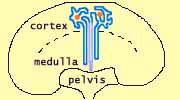 |
| This diagram is not drawn to scale. Each kidney comprises hundreds of thousands of
nephrons. |
A gross section of the kidney reveals that the outer cortex has a
somewhat different texture from the deeper medulla. This difference
reflects the disposition of various portions of the many nephrons
which comprise the kidney.
- The cortex consists of convoluted tubules
together with the renal corpuscles.
- The medulla consists of loops of Henle and
collecting ducts.
- The medulla may be divided into "zones"
or "stripes," visible grossly, which reflect the structural
differentiation of the tubules that form the loops of
Henle.
- The medulla has a remarkable interstitial environment,
hypertonic and poorly oxygenated. (Consult your physiology resources
for more info.)
- The cortex and medulla together comprise millions of individual nephrons,
all packed together.
- The cortex and medulla surround and drain into the hollow pelvis,
the funnel-shaped beginning of the ureter. Like
the ureter, the pelvis is lined by transitional
epithelium.
|
renal cortex
|
renal medulla
|
renal pelvis
|
 |
 |
 |
 The cortex / medulla organization has considerable functional significance, reflected in the arrangement of renal
tubules and vasculature.
TOP
OF THIS PAGE / CONTENTS
FOR THIS PAGE / RENAL IMAGE INDEX
Lobes and lobules
(Renal lobes and lobules are details, of little clinical significance. Awareness of their existence does help explain
some of the terminology for describing renal vasculature. Their existence also reflects embryonic development.)
Each kidney lobe consists of a medullary pyramid (a roughly pyramidal region that projects into the pelvis)
and its associated cortex. Lobes are visible to gross
inspection of a sectioned kidney. Medullary pyramids are separated from one another by cortical tissue
that extends deeper into the kidney, the so-called "columns of Bertin" (named after E.-J. Bertin, b. 1712).
A renal lobule is defined as a portion of the kidney containing those
nephrons that are served by a common collecting
duct.
Lobules are generally inconspicuous, even to microscopic
inspection.
Lobules are centered on "medullary rays,"
bundles of straight tubules (collecting ducts
and loops of Henle) which resemble the substance of
the medulla but extend into the cortex.
 A
complementary grouping of nephrons is provided by the
distribution of afferent arterioles from a common
interlobular artery. The interlobular
artery ascends through the cortex between lobules
and sends out afferent arterioles to nearby glomeruli.
TOP
OF THIS PAGE / CONTENTS
FOR THIS PAGE / RENAL IMAGE INDEX
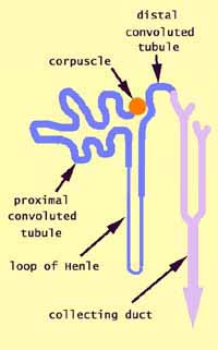
The NEPHRON
The nephron is the functional unit of the kidney. Each
nephron consists of one renal corpuscle and its associated
tubule. The kidney as a whole consists of many
nephrons (millions) with their associated blood vessels.
The renal corpuscles are the sites where
the process of urine formation begins with a filtrate of blood plasma.
Renal tubules are differentiated into several
segments. In the diagram to right, click on any tubule segment for more detailed information.
Each of these nephron components is described in greater detail below.
TOP
OF THIS PAGE / CONTENTS
FOR THIS PAGE / RENAL IMAGE INDEX
|
The RENAL CORPUSCLE
The most distinctive microscopic feature of the kidney is the
renal corpuscle. Renal corpuscles produce a filtrate of blood
plasma. (For more on this glomerular function, see below.
Also see CLINICAL
NOTE, above.)
Historical note: Renal corpuscles may also be called "Malpighian
corpuscles," after Marcello Malpighi (b. 1628), the researcher
who introduced microscopy to medicine.
|
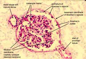
Click on image for enlargement.
|
|
|
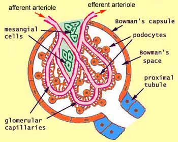
Each renal corpuscle has several parts. In the diagram to right, click on any feature for
more detailed information, or continue down the page.
- Bowman's capsule is the outer, epithelial
wall of the corpuscle.
- Bowman's space, also called "urinary
space," is the space (white in the diagram) lying within Bowman's capsule.
- The glomerulus, comprising several
distinct elements, is the conspicuous "little
knot" which occupies most of the corpuscle.
- Glomerular capillaries
have an endothelium that is fenestrated (full of holes).
- Podocytes are epithelial
cells covering the glomerular capillaries.
- Immediately adjacent to each glomerular capillary,
between the podocytes and the capillary endothelium, is the
filtration membrane (unlabelled red line on this
diagram).
- Mesangium is a supporting
tissue consisting of mesangial cells and matrix (green in the diagram).
- The beginning of the proximal tubule (blue in the diagram) is
the "drain" carrying fluid away from Bowman's space.
|
|
Each of these glomerular components is described in greater detail below.
TOP
OF THIS PAGE / CONTENTS
FOR THIS PAGE / RENAL IMAGE INDEX
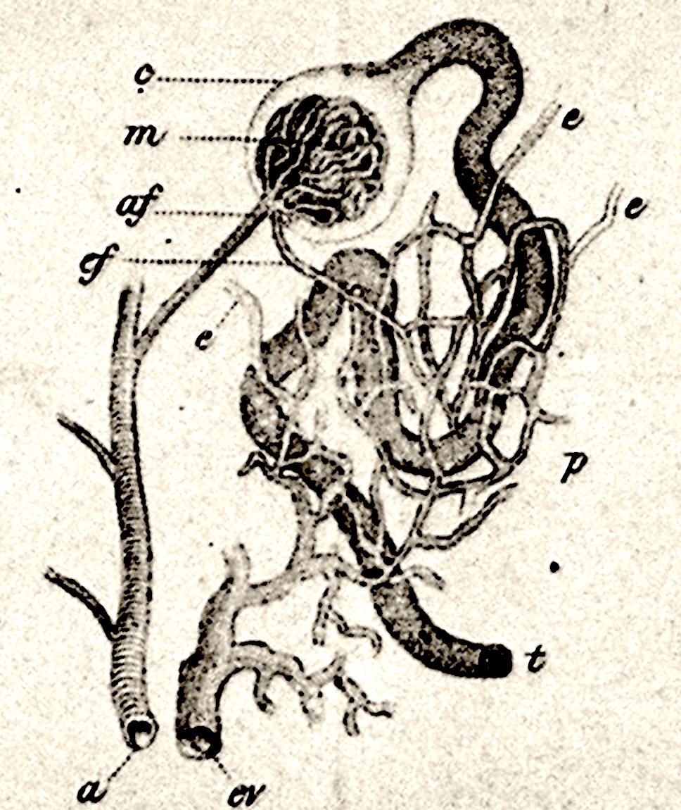
BOWMAN'S SPACE and BOWMAN'S CAPSULE
Historical note: Bowman's capsule and Bowman's space
are both named after English surgeon William Bowman.
At right is an image of Bowman's 1842 diagrammatic drawing of basic renal cortical structure.
Click on this image for additional historical information.
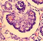
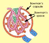 Bowman's space, also called the urinary space, is the space within Bowman's
capsule surrounding the loops and lobules of the glomerulus.
This is the space into which the glomerular plasma filtrate collects as it
leaves the capillaries through the filtration membrane.
Bowman's capsule is the outer epithelium which encloses Bowman's
space. This epithelium is simple
squamous, becoming cuboidal at the proximal tubule.
 Although
Bowman's capsule is rather obviously a simple
squamous epithelium, it is less apparent that the glomerulus
is also closely enveloped by epithelium. The peculiar structure of podocytes
obscures the fact that they are indeed epithelial cells. Thus Bowman's
space is entirely lined by epithelium.

The outer, parietal epithelium of the renal corpuscle is Bowman's capsule.
The inner, visceral epithelium is comprised of podocytes.
One way to appreciate the essential epithelial construction of renal corpuscles
is by examining fetal development. Click
on diagram or image to view a series of micrographs of developing renal corpuscles.
 Each renal
corpuscle is roughly spherical and has two "poles," not necessarily at opposite
ends of the corpuscle.
A random section across a renal corpuscle will only rarely display either pole. Yet, as you might
have noticed, images of renal corpuscles in this website (and elsewhere) commonly include both
poles. These micrographs were taken during hours of searching across various slides, with examination
of many, many corpuscles, to find those which had been sliced through both poles by a fortuitous plane of section.
TOP
OF THIS PAGE / CONTENTS
FOR THIS PAGE / RENAL IMAGE INDEX.
|
The GLOMERULUS
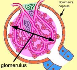
 The glomerulus ("little ball of yarn") is essentially a small
knot of capillaries and supporting structures suspended within Bowman's
capsule. The glomerulus is the source of the initial filtrate
of plasma that is eventually processed into urine. With this function,
the glomerulus is arguably the most significant component of the nephron.
|
 |
|
Several elements comprise the glomerulus. Click on any feature
for more detailed information, or continue down the page.
- Cells
- Extracellular materials
|

|

 In typical histological specimens, it is often difficult to discern the relationships
among these elements (i.e., the glomerulus looks like a jumbled mass of cells). Very thin sections (2µ or less) are much better
than routine thick (5-10µm) sections for resolving cell types and their relative positions.
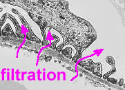
 Electron
microscopy is essential for resolving functional details
like capillary fenestrations and podocyte filtration slits (consult your histology
text or atlas, e.g., Rhodin figures 32-9, 32-10, 32-11).
|
|
|
|
|
|
|
Several pathological processes modify glomerular structure
and thereby interfere with glomerular filtration. Diagnosis of renal pathology routinely calls
for histological examination of glomerular details, including electron microscopy. See examples
from WebPath:
- "Minimal
change disease (MCD) with effacement of podocyte foot processes."
Note that podocytes form a continuous surface over the capillary; filtration
slits are missing.
- "Membranous
glomerulonephritis with thickened glomerular basement membrane containing
electron dense deposits."
Glomerular capillary endothelium
Introductory note about
capillaries.
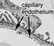 |
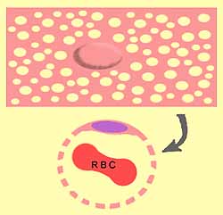
The endothelium of glomerular capillaries is perforated
with many small holes, or fenestrations (from Latin fenestra,
window). Each fenestrated endothelial cell has a shape like a slice of very
holey Swiss cheese, rolled into a cylinder to make a segment of capillary.
The fenestrations are too small to allow blood cells through, but plasma
can pass freely out of the holes and into the filtration
membrane.
The capillaries of renal glomeruli are exceptionally
leaky. Although the filtration membrane
holds back cells and plasma proteins, the remaining fluid (water, mineral
ions, and small molecules) passes freely into Bowman's
space and hence along the renal tubule.
Consult your histology textbook and/or atlas (e.g.,
Rhodin, figure 32-9 , 32-10, and 32-11) for additional
detail and electron micrographs of these cells.
|
 |
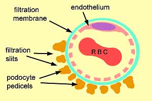
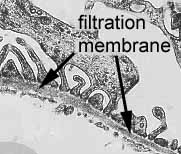
Filtration membrane
Immediately outside the capillary endothelium is the filtration
membrane (blue-greenish in the diagram to right). This membrane represents a fusion of the endothelial
basement membrane with
the basement membrane
of the glomerular epithelium (the podocytes).
Pathology: Anything
which clogs or thickens the filtration membrane can interfere with
the passage of fluid and hence reduce the rate of filtration.
See WebPath.
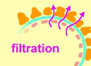
As plasma passes through the capillary fenestrations,
water, ions, and small molecules pass freely through the filtration membrane
into Bowman's space, while serum proteins are
retained in the capillaries. The outside of the filtration membrane
is supported by podocytes.
|
 |
 |
The filtrate which accumulates in Bowman's space drains into the proximal
tubule, and hence to the loop of Henle,
the distal tubule, and the collecting
duct. In these various segments of the renal
tubule, the filtrate is modified into urine, chiefly by reabsorption
of non-waste components.


The filtration membrane is not apparent
on H&E stained histological
specimens but may be demonstrated with PAS
or silver stain. Electron
microscopy is the best way to visualize the filtration membrane.
|
Podocytes
|
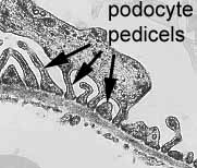
Podocytes ("footed cells") are extraordinary (one
might say bizarre) epithelial cells which support the filtration
membrane without obstructing the flow of filtrate. Each podocyte
stands upon branched pedicels, or "foot processes," which
rest on the filtration membrane. Between
adjacent pedicels are gaps called filtration slits which permit
free passage of fluid filtrate into Bowman's space.
In mature renal corpuscles, the epithelial nature of podocytes is not obvious.
Nevertheless, observation of fetal
development reveals that podocytes form from the epithelial lining
of Bowman's capsule.
 In typical histological preparations, podocyte nuclei tend to be oval and
fairly euchromatic. They may be recognized because they look more
"epithelial" than those of capillary
endothelial cells or mesangial cells, and
because they lie immediately adjacent to Bowman's
space (no intervening tissue).
Consult your histology textbook and/or atlas (e.g.,
Rhodin, figure 32-9 , 32-10, and 32-11) for additional
detail and electron micrographs of these cells.
Pathology: Anything
which occludes filtration slits or Bowman's space
can interfere with the passage of fluid and hence reduce the rate
of filtration.
Fusion of adjacent pedicels can block filtration
(e.g., see WebPath),
as can occlusion of Bowman's space caused by
proliferation of any of the cell types of the renal corpuscle (podocytes,
mesangial cells, Bowman's capsule cells, capillary endothelial cells).
|
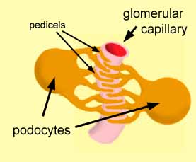 |
This simplified diagram is not drawn to scale, and does not
show the filtration membrane.
|
|
 Mesangial cells and matrix
Glomerular mesangial cells are inconspicuous and rather non-descript
cells concentrated toward the vascular pole of the glomerulus.
These cells produce the mesangial matrix and may contribute to maintenance
of the filtration membrane.
Mesangial cell nuclei may sometimes be recognized as small,
irregularly shaped, and rather heterochromatic nuclei within the glomerulus.
Consult your histology textbook and/or atlas (e.g., Rhodin,
figure 32-9 and 32-10) for additional detail and electron
micrographs of these cells.
Exra-glomerular mesangial cells, also called lacis cells or cells
of Goormaghtigh (commemorating Norbert Goormaghtigh, b. 1890) are
found in the space between the glomerulus and the macula
densa of the distal tubule; they form part of the juxtaglomerular apparatus.
The mesangial matrix is extracellular material which surrounds the
mesangial cells. Apart from offering some mechanical support to the
glomerular capillaries, the function of mesangial matrix is unknown.

 The mesangial matrix is not apparent on H&E
stained histological specimens, but (like the filtration
membrane) it may be visualized with PAS
or silver stain.
TOP
OF THIS PAGE / CONTENTS
FOR THIS PAGE / RENAL IMAGE INDEX.
RENAL TUBULES
In each nephron, the renal tubule receives plasma filtrate draining from Bowman's space
and processes it into urine.
- Each tubule is differentiated into several specialized segments.
- Different aspects of filtrate reabsorption are localized in different
segments.
The functional differentiation of the different tubular segments is associated
with variation in the structure of tubular epithelial cells. This structural
specialization is in turn reflected in the microscopic appearance of the tubules.
Click on any feature for more detailed information, or
continue down the page, where each segment is described further.
|
|
 |
|
Proximal convoluted tubule
The initial segment of the tubule is the proximal convoluted tubule.
It is called proximal because it is nearest to the starting point
(the renal corpuscle) and convoluted
because it twists about (in contrast to the straight segments of tubule
which form the loop of Henle). This segment
of the renal tubule restores much of the filtrate to the blood in the
peritubular capillaries, by actively pumping
small molecules out of the tubule lumen into the interstitial space.
Water then follows the concentration gradient.
The length of a proximal convoluted tubule tends to be several times
greater than that of a distal convoluted tubule,
so sections of proximal tubules are several times more numerous than those of distal
tubules in a typical histological slide of renal cortex.
The epithelium of the initial descending segment of the loop
of Henle is similar to that of the proximal convoluted tubule, and
is sometimes called the pars recta of the proximal tubule (in
contrast to the convoluted pars convoluta).
|
 |
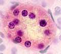 |
The proximal convoluted tubule is lined by a simple
cuboidal epithelium whose cells have several characteristic features, each of which affects the appearance of the tubule.
- The apical end of each proximal tubule cell has a brush border of
microvilli. This provides an increased surface area to accommodate
the membrane channels that are responsible for absorbing into the
cell small molecules from the filtrate in the tubular lumen.
- Appearance: The brush border is seldom plainly visible in
routine histological preparations, but proximal tubule cells tend
to have indistinct apical ends (in contrast to the more definite
apical border of cells comprising distal tubules
and collecting ducts).
- Proximal tubule cells have a high proportion of mitochondria
in their cytoplasm, to provide the energy for pumping ions and molecules
against their concentration gradient.
- Appearance: The abundance of mitochondria
makes proximal tubule cells
rather intensely acidophilic.
- The plasma membranes of adjacent proximal tubule
cells are extensively interdigitated. This increases the basal
membrane surface area available for pumping molecules out the basal
end of each cell.
- Appearance: As a consequence of such interdigitated cell
membranes, boundaries between adjacent proximal tubule cells are
inconspicuous (i.e., in section the epithelium looks like a continuous
band of cytoplasm with nuclei appearing at irregular intervals).
Consult your histology textbook and/or atlas (e.g.,
Rhodin, figure 32-13 and 32-14) for additional detail
and electron micrographs of these cells.
|
 Loops
of Henle (medullary tubules)
The loop of Henle is a remarkable feature of the renal tubule, associated
with the remarkable function of the renal medulla in
water conservation. Basically, the loop helps to establish and maintain a hypertonic
saline environment in the medulla, via a counter-current exchange process, which in turn
allows subsequent recovery of water from collecting
ducts (and associated concentration of urine within the collecting
ducts). For further explanation of this process, see your physiology
resources.
Historical note: The loop of Henle is named for Friedrich Henle, b. 1809.
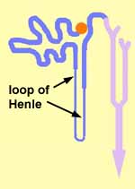
The loop of Henle consists of a descending limb, having an
initial short thick segment followed by a long thin segment, and an ascending
limb, having a thin segment followed by a thick segment.
|
Distal convoluted tubule
The distal convoluted tubule continues into the cortex from
the ascending limb of the loop of Henle. Like
the proximal convoluted tubule, this segment of the tubule is called
convoluted because it twists about. It is distal
because it is further "downstream" from the starting point,
the renal corpuscle.
Like the proximal convoluted tubule, this distal segment of
the renal tubule continues the return of useful materials from the filtrate
to the blood in the peritubular capillaries, by actively
pumping small molecules out of the tubule lumen into the interstitial
space.

 Each distal tubule returns to the vascular pole of the
renal corpuscle from which the tubule arose.
At this site is a special region of the distal tubule called the macula
densa (macula densa = "dense spot," named for the
close clustering of epithelial nuclei in the wall of the distal tubule)
which is part of the juxtaglomerular apparatus.
In typical histological specimens of renal cortex, profiles of distal convoluted tubules tend to be relatively less numerous
than those of proximal convoluted tubules, because distal tubules
tend to be considerably shorter than proximal tubules.
|
 |
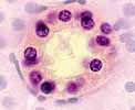 |
The distal convoluted tubule is lined by a simple
cuboidal epithelium whose cells have several characteristic features, each of which affects the appearance of the tubule.
- Unlike the proximal convoluted
tubule, the apical end of each distal tubule cell does not have
a brush border, although there may be scattered microvilli.
Because most of the "heavy lifting" has already been done
in the proximal tubule, distal tubule cells
are not so highly specialized.
- Appearance: The apical ends of distal tubule cells tend
to be more distinct than those of proximal
tubule cells, conferring the usual appearance of a larger,
clearer lumen in each distal tubule.
- Like proximal tubule cells, distal tubule cells have a high proportion of mitochondria
in their cytoplasm, to provide the energy for pumping ions and molecules
against their concentration gradient.
- Appearance: However, distal tubule cells are less extremely
specialized than those of the proximal tubules.
They have relatively fewer mitochondria than proximal tubule cells and therefore display a lesser degree of
acidophilia.
- The plasma membranes of adjacent distal tubule cells
are extensively interdigitated (as are those of proximal
tubules). This increases the basal membrane surface area
available for pumping molecules out the basal end of each cell.
- Appearance: As a consequence of such interdigitated cell
membranes, boundaries between adjacent distal tubule cells are
inconspicuous (i.e., in section the epithelium looks like a continuous
band of cytoplasm with nuclei appearing at irregular intervals).
Consult your histology textbook and/or atlas (e.g.,
Rhodin, figure 32-19 and 32-20) for additional detail
and electron micrographs of these cells.
|
TOP
OF THIS PAGE / CONTENTS
FOR THIS PAGE / RENAL IMAGE INDEX
Macula densa / juxtaglomerular apparatus (juxtaglomerular
complex)
|
|
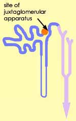
The juxtaglomerular apparatus is a complex of structures associated
with the vascular pole of each renal corpuscle.
The juxtaglomerular apparatus has two principal components:
- The macula densa is a patch of densely-packed epithelial
cell nuclei along the distal convoluted tubule,
adjacent to the afferent arteriole at the
vascular pole of the corpuscle from which
the tubule arose. It may function as a sensor for sodium and/or
chloride concentration.
- Juxtaglomerular cells ("J-G cells") in the wall
of the afferent arteriole are specialized
smooth muscle cells containing secretory granules, the source of the
hormone renin.
- Consult your histology textbook and/or atlas
(e.g., Rhodin, figure 32-6 and 32-7) for additional
detail and electron micrographs of these cells.
- The juxtaglomerular region also includes extra-glomerular mesangial
cells, also called cells of Goormaghtigh (commemorating
Norbert
Goormaghtigh, b, 1890), or lacis cells.
The juxtaglomerular apparatus participates in the
regulation of blood pressure and the rate blood flow through the glomerular capillaries (and hence
the rate of urine formation).
|
|
Understanding the detailed histophysiology of the juxtaglomerular apparatus has been a principal
achievement of modern nephrology. For a detailed historical account of this understanding, see
"The juxtaglomerular
apparatus of Norbert Goormaghtigh -- a critical appraisal," by G. Eknoyan, et al. (2009) Nephrology
Dialysis Transplantation, V. 24, pp. 3876-3881 (https://doi.org/10.1093/ndt/gfp503).
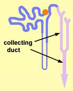 Collecting
ducts
Collecting ducts are named because they "collect" the urine
from distal tubules.
Larger collecting ducts may also be called papillary ducts or ducts of Bellini.
Historical note: The name "ducts of Bellini" commemorates Lorenzo Bellini, the 17th
century Italian anatomist who first described them.
Collecting ducts are lined by simple cuboidal epithelium which appears
less specialized than that of the proximal or distal
tubules. Cytoplasm of collecting duct cells is relatively clear (i.e., not as intensely
eosinophilic as that of proximal or distal tubules), and cell borders are usually distinct. Collecting ducts
merge and become larger as they descend through the medulla,
so different sizes of collecting ducts may be observed at different levels
in the kidney, with the smallest in the cortex and the largest near the pelvis.
- Collecting duct epithelium has the unusual physiological property of adjustable
permeability to water (under control of the pituitary
anti-diuretic hormone, ADH). If permeability to water is high,
then water diffuses across the collecting duct epithelium into the hypertonic
interstitium of the medulla, resulting in a concentration of urine within
the duct. But if water permeability is low, then water is retained
in the urine and excreted from the body.
Collecting ducts are readily recognized in the renal medulla, as relatively
large tubules lined by cuboidal epithelium, in which the epithelial cells
are relatively clear (i.e., not as eosinophilic as proximal and distal tubules)
and have distinct cell borders.
TOP
OF THIS PAGE / CONTENTS
FOR THIS PAGE / RENAL IMAGE INDEX
 RENAL VASCULATURE
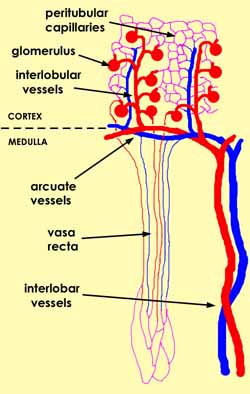
 In most organs the particular disposition of arteries, veins and capillaries
serves only to distribute blood uniformly throughout the organ. In the
kidney, however, the arrangement of blood vessels has special functional significance.
Here we will offer a more-detailed-than-usual account of the within-organ
arrangement of blood vessels. (Click here
for a general introduction to blood vessels.)
|
Distributing vessels.
See clinical note for the surgical significance
of renal vessel arrangement.
- Interlobar arteries and veins arise from the renal
artery and vein and ascend between lobes (as the adjective interlobar
suggests) from the pelvis across the medulla
toward the cortex.
- Arcuate arteries and veins branch from the interlobar
vessels and "arch" (as the adjective arcuate suggests)
across the boundary between cortex and medulla.
CLINICAL NOTE: Interlobar and
arcuate vessels do not extend into the cortex. Therefore, to
minimize the risk of bleeding during renal biopsy, the biopsy needle
should be aimed tangentially through the cortex to avoid damaging
one of these larger vessels.
- Interlobular arteries and veins ascend from the arcuate
vessels and pass up into the cortex perpendicular
to the surface of the kidney. These may also be called cortical
radial vessels. "Radial" describes their
orientation in the cortex. "Interlobular" refers to
renal lobules, which are an inconspicuous pattern of organization for renal cortex.)
 In
some of our kidney specimens, the distributing vessels display conspicuous
atherosclerosis. In
some of our kidney specimens, the distributing vessels display conspicuous
atherosclerosis.
|
|
|
Microvasculature. The kidney has
three distinct capillary networks, each with a different function.
|
|
|
 |
|
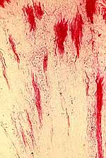
Vasa recta
|
|
|
TOP
OF THIS PAGE / CONTENTS
FOR THIS PAGE / RENAL IMAGE INDEX
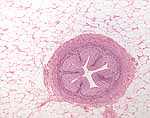
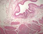
Urinary Tract
The tissue composition of renal pelvis, ureters, bladder and urethra
is relatively simple, compared to kidney. Each of these elements has a wall lined by
transitional epithelium (urothelium)
and containing smooth muscle.
The male urethra is associated with the prostate as well as with
bulbourethral glands and periurethral glands.
The female urethra is associated with paraurethral glands of Skene.
|
renal pelvis
|
ureter
|
bladder
|
urethra
|
 |
 |
 |
 |
 |
 |
TOF THIS PAGE / CONTENTS
FOR THIS PAGE / RENAL IMAGE INDEX
Renal Image Index
TOP
OF THIS PAGE / CONTENTS
FOR THIS PAGE
 Comments
and questions: dgking@siu.edu
SIUC / School
of Medicine / Anatomy / David
King
https://histology.siu.edu/crr/rnguide.htm
Last updated: 12 November 2025 / dgk
 Histology Study Guide

 The thumbnail to right links to some images of membranoproliferative
glomerulonephritis.


 Bowman's space, also called the urinary space, is the space within Bowman's
capsule surrounding the loops and lobules of the

 Each renal
corpuscle is roughly spherical and has two "poles," not necessarily at opposite
ends of the corpuscle.




 Loops
of Henle (medullary tubules)


 Collecting
ducts


 Comments
and questions: