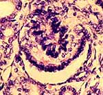
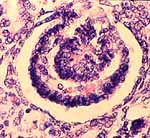
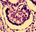
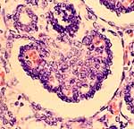
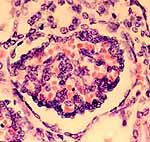
Developing renal corpuscles, fetal kidney
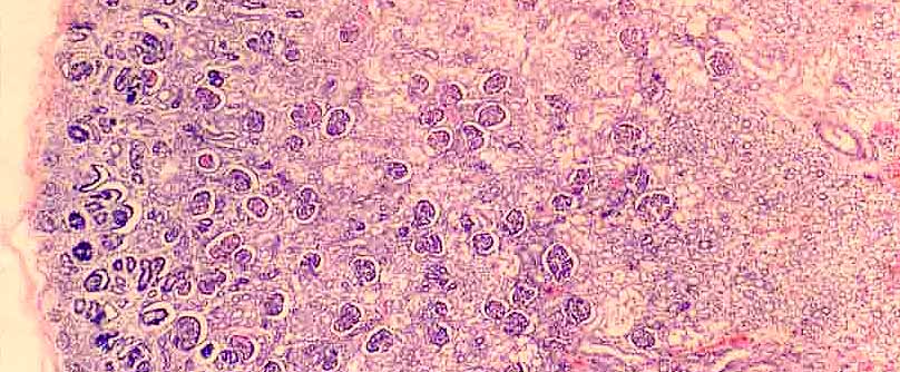
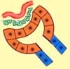
This specimen displays a range of stages of corpuscle development, progressing from left to right in the image above. As the developing kidney grows, new renal corpuscles are added near the surface of the cortex while those found deeper in the cortex formed earlier and are more mature.
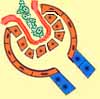
1. The renal corpuscle first appears as a curved vesicle. The adjacent condensation of mesenchyme is the precursor of the glomerulus. 2. A cuboidal epithelium covers the incipient glomerulus, while a squamous epithelium forms the outer lining of Bowman's capsule.
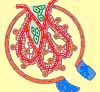
3. While podocytes differentiate over the developing glomerular capillaries, leftover mesenchyme (green) becomes the mesangium.
Comments and questions: dgking@siu.edu
SIUC / School
of Medicine / Anatomy / David
King
https://histology.siu.edu/crr/RN067b.htm
Last updated: 15 September 2021 / dgk