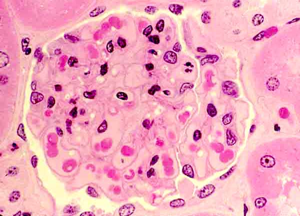

Renal corpuscle, thin section
 |
This 1µm section provides higher resolution than a routine 5-10µm section.
In this specimen, the various glomerular cell types can be distinguished by their relationships with capillary lumens, Bowman's space, and the filtration membrane, as well as by the appearances of their nuclei..
Cell identification (see below for a labelled image):
- Most glomerular capillaries contain red blood cells (RBCs). Glomerular capillary endothelial cells are locate on the capillary lumen side of the filtration membrane (i.e., across the membrane from Bowman's space). Endothelial nuclei are flattened (so their appearance depends on the plane of section) and fairly heterochromatic.
- Bowman's space appears relatively clear. (The small clear "bubbles" within capillaries are artefacts.)
- Podocyte cell bodies are located in Bowman's space. Their nuclei are relatively large and euchromatic.
- Mesangial cells are surrounded by mesangial matrix and glomerular capillaries. Mesangial cell nuclei are relatively small, irregular in shape, and heterochromatic.
RENAL IMAGE INDEX
Comments and questions: dgking@siu.edu
SIUC / School
of Medicine / Anatomy / David
King
https://histology.siu.edu/crr/RN109b.htm
Last updated: 16 September 2021 / dgk