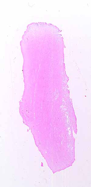
This is a fairly difficult specimen. The source
will not be obvious. Identification will require noticing some rather
subtle hints.
What kinds of tissue comprise the bulk of this specimen?
Examine (carefully!) the entire circumference of this specimen.
- How many distinct surface appearances can you find?
- Where do you find cut surfaces?
- How much of the circumference is lined by epithelium? (Note
that post-mortem specimens commonly shed epithelial cells from exposed
mucosal surfaces.)
- How many different kinds of epithelium can you find? Where?
- In what locations of the body might such epithelial surfaces be
found?
Notice the tissue composition, including appearance of blood vessels,
beneath the surface epithelium in each region of this specimen.
- Is there any glandular tissue? If so, describe the glands.
- Are any of the locations listed above also consistent with the deeper
tissues observed on this specimen?
Hints on the next page are a bit more pointed. Don't look
unless you are stuck.
More hints.