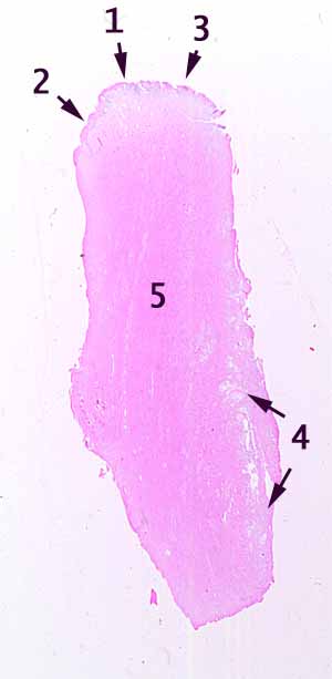
Find the region labelled 1 on the image.Note the types of epithelium which occur on either side, toward numbers 2 and 1.
Toward 3, the epithelium is poorly preserved. You may need to hunt a bit to find some patches of poorly attached epithelial cells. Post-mortem specimens commonly shed epithelial cells from exposed mucosal surfaces.)
Note the quality of connective tissue and vasculature underlying numbers 2 and 3. In which region are lymphocytes more numerous?
So, point 1 marks a transition in the composition of the mucosa. List all regions of the body where such a transition might occur. (If your list is complete, this specimen must have come from one of them.)
You should be able to find glandular tissue in the region indicated by 4.
Describe the cells which comprise these glands. In what part(s) of the body do glands such as these occur?
Finally, describe the tissue in region 5.
What region or regions of the body have all of the features listed above?
Hints on the next page are quite revealing. Don't look until you are ready to give up.
