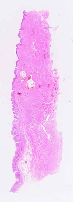
The source for this specimen should be obvious (and confidently
so), provided you are familiar with the organ it represents and don't
overlook any conspicuous clues.
Note the various tissue layers.
- Name these layers.
- Are the layers similar along the entire length of the specimen?
Which organs have such distinct layers?
Note specific features of the mucosal layer.
- Make sure you examine the entire epithelial surface.
- What shape is the surface?
- How many different types of epithelium can you find?
- What kind(s) of cells comprise each type of surface epithelium?
- Are there any epithelial features (e.g., glands or crypts) embedded
in the lamina propria?
- Can you see a distinct muscularis mucosae? If so, does it
extend along the entire length of the specimen?
Are there any distinctive features in the submucosa?
Is the muscularis externa organized into distinct layers? What
kind of muscle comprises the muscularis in this specimen?
What are the bright red patches in the deeper tissues?
Identifying this slide should be easy. But be careful because
one incorrect identification may also be too easy.
Hints on the next page are quite revealing. Don't look until
you are ready to confirm your identification.