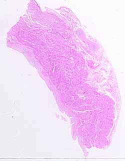
This may be one of the more difficult specimens in the SAQ series. There is, however, one small region that offers a reliable clue for locating the source.Note the various tissue layers.
Note specific features of the mucosal layer.
- For most hollow organs, the appearance of mucosal epithelium provides much information.
- Unfortunately, in this specimen, most of the surface epithelium is missing.
- Nevertheless, try to find as much of the epithelium as you can. (Preserved epithelium may be found within occasional folds of the mucosa.) Then try to imagine what types of epithelia might be consistent with these remnants.
- Also note the overall shape of the surface. Are there crypts, or villi, or other such special surface shapes?
Now note the muscularis. Is the muscularis organized into distinct layers? What kind of muscle comprises the muscularis in this specimen?
Does the outer (deepest) layer appear to be a serosa or an adventitia? (That is, does it display a smooth natural surface, or connective tissue that has been cut?)
Now, what organs are consistent with the above observations?
- You should be able to imagine several organs, in each of several organ systems, with smooth muscle in their walls.
- Some of these may be excluded on the basis of mucosal shape
- Others may be excluded on the basis of epithelium inconsistent with the visible remnants on this specimen.
But hidden somewhere on this specimen is an (unexpected?) clue which, if noticed and recognized, will reveal the location.
Hints on the next page are quite revealing. Don't look until you are ready to confirm your identification.
