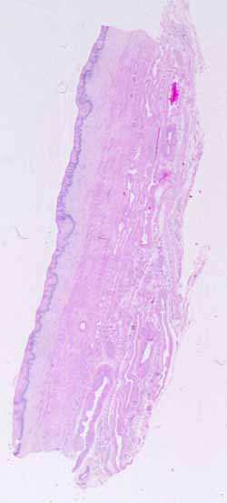
Hopefully, you have recognized that the mucosal surface of this specimen is lined by a nonkeratinized, stratified squamous epithelium. There are only a limited number of sites in the human body which have such an epithelium.
Next, hopefully, you have already noticed that the deeper tissues include considerable smooth muscle. This rules out the oral cavity (where all the muscle is striated).
Next, you may have noticed that there is no clearly defined submucosa and muscularis, nor distinct layers of circular and longitudinal muscle. Rather, deep to the lamina propria, many small bundles of smooth muscle are thoroughly interwoven with connective tissue throughout the wall. These features are inconsistent with esophagus, which (like the rest of the GI tract) has very well-defined layers of mucosa, submucosa, and muscularis.
Above are listed "negative" hints, ruling out possibilities. Positive hints include the following:
- Very extensive vasculature, including noticeable veins quite close to the surface (unlike skin).
- Extensive innervation, with lots of conspicuous nerves. (If you can't find or recognize any nerves, please ask your instructor for assistance. Several slides from this specimen include a parasympathetic ganglion.)
- Very pale cytoplasm (appearing "empty") in cells of the stratified epithelium.
No more hints.
