
Esophagus
The esophagus provides passage from the oral cavity to the stomach. Compared to other regions of the GI tract, the esophagus is a fairly simple tube. Functional specializations are correspondingly few.
The esophageal lining is protected by a stratified squamous epithelium. Because this epithelium is normally not exposed to dryness or to abrasion, it is non-keratinized.
Scattered submucosal mucous glands provide lubrication.
A well-developed muscularis provides peristaltic propulsion of food. (Even when upside down, the esophagus can push food and drink to the stomach.)
The basic layers of the GI tract are especially distinct in the esophagus, in contrast to the vagina.
In the esophageal mucosa:
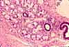
Esophageal epithelium is non-keratinized stratified squamous. Note that the basal surface of the epithelium can be deeply indented by connective tissue papillae.
In oblique section through the epithelium, the connective tissue papillae can look like "islands," apparently surrounded by epithelium. Beginning students frequently mistake these for glandular structures.
Epithelial continuity is critical for normal function. A breach in the epithelium creates an ulcer. For an image of an esophageal ulcer, see WebPath or Milikowski & Berman's Color Atlas of Basic Histopathology, pp. 162-163.
Esophageal epithelium may be transformed to a simple columnar form in the condition called Barrett's esophagus. The epithelium is variously described as resembling that of gastric mucosa (i.e., with tubular glands) or of intestinal mucosa (i.e., with goblet cells). The cause of this condition remains uncertain, but it may represent a metaplastic response to chronic inflammation (caused, e.g., by gastric reflux). Barrett's esophagus can be associated with esophageal obstruction from scarring and/or carcinoma. (For images, go to WebPath and/or see Milikowski & Berman's Color Atlas of Basic Histopathology, pp. 234-236.)
At the junction with the stomach, the stratified squamous epithelium of the esophagus makes an abrupt transition to the simple columnar epithelium of the gastric mucosa.
Esophageal lamina propria is less cellular (fewer lymphocytes) than lamina propria in the stomach and intestine, presumably because the protective stratified squamous epithelium is more effective at keeping out foreign antigens.
Lymph nodules are uncommon in the esophagus, but they may occur here occasionally, as elsewhere throughout the GI tract.
Esophageal muscularis mucosa is noticably thicker than that in the stomach and intestine, and includes only longitudinal muscle fibers.
(Because the longitudinal fibers occur in bundles, a longitudinal section passing between bundles may not include any evidence of muscularis mucosae.)
Connective tissue of the esophageal submucosa is typically more fibrous and less cellular than that in the lamina propria of the mucosa.
Small mucous glands are scattered in the esophageal submucosa. Ducts from these glands pass through the surface epithelium to drain into the esophageal lumen. Within the submucosa the ducts branch into tubular secretory units which are lined by mucus-secreting cells. These glands provide mucus for lubricating the passage of food down the esophagus, augmenting the role of salivary glands.
In the esophagus, the submucosal vascular plexus includes especially large venous spaces. These may enlarge into esophageal varices, especially in cases of portal hypertension (an increase in pressure in the portal vein, due to cirrhosis). Such varices carry a substantial risk of rupture with fatal bleeding into the esophageal lumen. (For more, go to WebPath or see Robbins Pathologic Basis of Disease.)
Excessive development of collagen in the submucosa, as in scleroderma, can cause a reduction in esophageal motility (see WebPath, scleroderma).
The muscularis externa of the esophagus consists of the standard inner circular and outer longitudinal layers of smooth muscle, with Auerbach's plexus in between.
In the upper third of the esophagus, the muscularis consists of striated muscle.
In the lower two-thirds of the esophagus, the muscularis is smooth muscle.
In the transitional region (thumbnails to right), both types of muscle may be seen.
The vagina provides an instructive contrast with the esophagus. The vagina and the esophagus are both tubular organs with a mucosa lined by non-keratinized stratified squamous epithelium. Hence these two organs appear superficially similar. However, bundles of smooth muscle are interwoven with connective tissue throughout the wall of the vagina, so that there is no clear demarcation of submucosal and muscularis layers.
Certain special features which distinguish these organs (e.g., abundance of glycogen storage in vaginal epithelium, submucosal glands in the esophagus) are not always apparent in routine sections. So the difference between "layers distinct" and "layers not distinct" may be the easiest and most reliable way to distinguish these two organs (e.g., on a practical evaluation).
Comments and questions: dgking@siu.edu
SIUC / School
of Medicine / Anatomy / David
King
https://histology.siu.edu/erg/esoph.htm
Last updated: 20 June 2023 / dgk