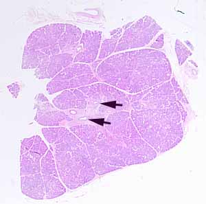
Hopefully, you have found ducts which identify this as an exocrine gland. If you hadn't recognized the large ducts in the interlobular connective tissue (arrows), look again now. (Make sure you distinguish ducts from blood vessels; the ducts are the ones with the cuboidal-to-columnar epithelium, two-layered in the larger examples.)
Hopefully, you have also recognized both serous acini and mucous tubules, identifying this as a mixed gland. If not, please examine the specimen again after reviewing the distinction between serous and mucous secretion. If you've gotten this far, don't worry about which particular mixed gland (as long as you can tell that is NEITHER parotid gland NOR pancreas).
Make sure you can distinguish between the secretory parenchyma (serous and mucous cells) and the numerous small intralobular ducts. (In small ducts, the nuclei are centrally located and the cytoplasm is not conspicuously polarized.)
Also notice the scattered patches of inflammatory infiltrate (regions of glandular stroma packed with small round nuclei of lymphocytes).
Note the number of adipocytes scattered throughout the stroma. (Trivia: Fat cells are common in intralobular stroma of salivary glands but rare in normal pancreas.)
No more hints.
