Glands of the Gastrointestinal System
Introduction to Glandular Tissue
( Basic Terminology)
Glands are organized arrangements of secretory cells. All exocrine
glands (and also most endocrine glands), are composed of
epithelial tissue. Although most glands
give the appearance of being "solid" tissue, their epithelial nature
is expressed by the organization of secretory cells into tubules,
acini, or cords. Every exocrine secretory cell
has some portion of its plasma membrane exposed to an external surface, communicating
with the outside of the body by a system of ducts.
Histologically, glands are described using some standard vocabulary, with
which you should be familiar.
Serous / Mucous / Mixed
 The
serous / mucous distinction
is based on the secretory cell's product -- whether it is a clear, watery
solution of enzymes (serous, like serum)
or else a glycoprotein mixture (mucous, like mucin).
These two categories of secretory products come from two distinct categories
of cells, each with a characteristic appearance.
The
serous / mucous distinction
is based on the secretory cell's product -- whether it is a clear, watery
solution of enzymes (serous, like serum)
or else a glycoprotein mixture (mucous, like mucin).
These two categories of secretory products come from two distinct categories
of cells, each with a characteristic appearance.

Mixed glands (e.g., most
salivary glands) contain both types of cells.
Glands which contain only one of these two cell types may be described
either as serous glands (e.g., parotid gland or
pancreas) or as mucous glands (e.g., Brunner's
glands).
Serous Secretion
 Serous cells are specialized to secrete
an enzyme solution. Examples include serous cells of the salivary
glands, exocrine cells of the pancreas, gastric
chief cells, and Paneth cells of intestinal
crypts. Serous cells of the pancreas
and the salivary glands are typically organized into secretory units called
acini.
In routine light microscopy, serous cells
are distinguished by basophilic basal cytoplasm, a centrally-located nucleus,
and variously-staining secretory vesicles (zymogen granules) in apical cytoplasm.
These features are all associated with organized mass production of
protein for export. More.
Mucous Secretion
 Cells
which are specialized to secrete mucus are called mucous cells. Examples
in the GI system include secretory cells of the salivary
glands, esophageal glands, stomach
surface, pyloric glands, and Brunner's
glands of the duodenum. These cells are typically organized into
tubular secretory units.
 Goblet
cells are mucous cells which stand apart from one another (between cells of other types) within a columnar epithelium.
Goblet cells take their name from their characteristic shape, with a
broad opening at the apical end and a narrow, "pinched" base. Cells
with this goblet shape are also characteristic of the respiratory tract
and the female reproductive tract.
Goblet
cells are mucous cells which stand apart from one another (between cells of other types) within a columnar epithelium.
Goblet cells take their name from their characteristic shape, with a
broad opening at the apical end and a narrow, "pinched" base. Cells
with this goblet shape are also characteristic of the respiratory tract
and the female reproductive tract.
In routine light microscopy, mucous cells
are most conspicuously distinguished by "empty"-appearing (i.e.,
poorly stained) apical cytoplasm and by densely-stained, basal nuclei. More.
Simple / Compound

The simple / compound distinction is based on on duct shape.
A simple
gland has either an unbranched duct or no duct at all. In either case,
there is only a single secretory unit (acinus or tubule).
Examples include sweat glands, individual gastric
glands, and intestinal crypts.

A compound gland has a branching duct. Salivary
glands and pancreas are familiar examples. Compound
glands are typically fairly bulky and contain very many individual secretory
units (acini or tubules).
Acinus / Tubule
/ Cord
Each secretory unit of a gland consists of cells arranged into an acinus,
a tubule, or a cord.
Each of of these arrangements has a different and characteristic appearance
when viewed in section.
Acinus (or alveolus)
 An
acinus (from Latin, grape; plural, acini) is a small ball of secretory epithelial
cells containing a tiny central lumen. Acini are usually formed by serous
cells. [Acini are sometimes called alveoli, from L., small
cavity.]
 A typical acinar cell is shaped like a pyramid. Its basal surface,
located at the periphery of the acinus, rests on the basement membrane separating
the acinus from the underlying stroma. Its lateral
surfaces (the sides of the pyramid) are attached to adjacent secretory
cells. Its apical surface is free and faces the acinar lumen,
which communicates by duct with the outside. The
acinar cell's cytoplasm is also visibly polarized, usually with basophilic
basal cytoplasm and variously-staining secretory granules concentrated in
apical cytoplasm. For more, see serous
cells.
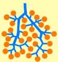
 A compound
acinar gland can be quite accurately modelled as a bunch of grapes
embedded in Jello™. The grapes are the acini, the branching stems
are the ducts, and the Jello™ represents the rest
of the stroma. Major and minor branches of the bunch
represent lobes and lobules, respectively, separated by greater amounts of
connective tissue.
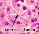
 In
routine tissue sections, most acini are cut in random planes and look like
solid lumps, made of cells having various sizes and shapes. The
lumen of an acinus is typically tiny (i.e., much smaller than a cell)
and so is visible only when an acinus is sliced neatly across the middle.
In such a slice, the cells look like slices of pie, with the lumen
in the center.
In
routine tissue sections, most acini are cut in random planes and look like
solid lumps, made of cells having various sizes and shapes. The
lumen of an acinus is typically tiny (i.e., much smaller than a cell)
and so is visible only when an acinus is sliced neatly across the middle.
In such a slice, the cells look like slices of pie, with the lumen
in the center.
Tubules

 In
contrast to the small balls of cells which comprise secretory acini,
secretory cells may also arrange themselves
into secretory tubules. This is a common form for mucous
glands (e.g., esophageal glands, pyloric
glands, Brunner's glands, mucous portions of salivary
glands). Other tubular glands include sweat glands and gastric
glands.
Because tubules are elongated, random sections commonly
include the lumen as well as the secretory cells themselves (in contrast
to the situation with acini). But interpretation
of the sectioned appearance of tubular glands will depend on whether the
tubules are simple or branched, on whether they are straight or twisted,
and on whether or not adjacent tubules lie parallel to one another.
Cords
 Cords
are arrangements of cells attached to one another to form sheets. In
section, the predominant pattern appears linear, even though the lines may
twist and branch.
Cords are a common arrangement for epithelial cells that are specialized
for endocrine secretion. The cells retain an epithelial
character, attached to neighboring cells, even though they may no longer comprise
a surface barrier between interstitial space and a secretory lumen that leads
to the outside. Examples of endocrine cells arranged into cords include
the epithelial cells of pancreatic islets, parathyroid,
adrenal cortex, and liver.
The liver is notable for having cells arranged
into cords in spite of its major exocrine function. In
order to maintain communication with ducts, the liver
cords contain a network of intercellular channels called bile
canaliculi.
Endocrine / Exocrine
The suffix -crine refers to secretion; the prefix endo- or
exo- tells where the secretory product goes.
The product of exocrine glands leaves the body proper, either by direct
secretion onto the body's surface (e.g., sweat) or into the lumen of an organ
(e.g., gastric juice) or else by flowing through a system of ducts
(e.g., saliva, pancreatic enzymes, bile). The cells of exocrine glands
are generally arranged into secretory units in the form of acini
or tubules (although the liver has a remarkable arrangement
of cords).
The product of endocrine glands is secreted into interstitial fluid
and hence into capillaries and general circulation. The cells of endocrine
glands are often arranged into cords adjacent to capillaries
or sinusoids.
Link to the endocrine
system.
Ducts
 Ducts
are relatively simple tubular structures which are (usually) easily distinguished
from blood vessels by their conspicuous cuboidal to columnar epithelial lining.
Blood vessels, of course, are lined by simple squamous endothelium.
The glandular cells which comprise ducts generally receive much less attention
than those which actually secrete the gland's product. However, the
complete understanding of a gland requires some awareness of and attention
to the duct system through which it drains. Ducts are not just passive "plumbing."
Some duct segments actively modify the secretory product passing through (typically
concentrating it by removing water).
In general, cells lining ducts may often be distinguished
by one or more of the following:

- cytoplasm rather pale (relative to most serous secretory
cells);
- nucleus centrally located (as opposed to somewhat basal
for most mucous secretory cells).
- basal and apical cytoplasm not obviously differentiated
(but this isn't always obvious for secretory cells either, and striated
ducts do have specialized basal cytoplasm);
- cells relatively short (cuboidal) relative to many (but
not all) secretory cells (and larger ducts may be lined by columnar cells);
For the purpose of describing duct structure and function,
some special terminology can be useful. However, by and large the distinctions
that these terms allow represent minor details rather than essential knowledge.
Intercalated / Striated Ducts
Intercalated ducts are small, short
ducts which drain individual secretory units. These are usually inconspicuous,
lined by a simple epithelium consisting of low cuboidal cells.
 Striated
ducts are duct segments specialized for concentrating the secretory product
that is flowing through the duct. Striated ducts are found in some but not all
glands (notably salivary glands), where they follow
after intercalated ducts and are lined by a simple epithelium consisting of
conspicuous cuboidal to columnar cells.
The cells of the striated ducts are specialized for concentrating
the secretory product that is flowing through the duct. They do this by pumping
water and ions across the duct epithelium, from the duct lumen and into
interstitial fluid. Remember the kidney?
An extreme example of the striated duct is represented by the proximal
tubule of a nephron in the
kidney.
Ultrastructurally, striated duct cells display extensive
infoldings of the basal membrane. These folds are closely associated
with mitochondria that provide ATP for the membrane pumps. In light
microscopy, the basal folds and mitochondria are sometimes visible as basal
striations, hence the name striated duct.
Secretory / Excretory Ducts

Both intercalated and striated ducts are sometimes called secretory ducts.
They are located within lobules (intralobular, next paragraph).
More distal ducts (interlobular, next paragraph), sometimes called
excretory ducts, are generally passive conducting tubes. Their
size varies, depending on how many branches have converged distally. Larger
excretory ducts may be lined by columnar or by stratified cuboidal
epithelium.
It is sometimes convenient to refer to ducts by location
within the gland. The following terms are all directly descriptive.
Intra- means within. Inter-
means between. Lobes and lobules are clusters of
secretory units served, respectively, by major and minor branches of the duct
tree. Within a lobule, individual secretory units are separated from
one another by little more than basement membranes and capillaries. In
contrast, the stroma which separates lobules and lobes consists
of thicker septa of connective tissue. (The distinction between lobes
and lobules is arbitrary; lobes are evident upon gross inspection while lobules
are evident to low power microscopy.)
 Intralobular
-- Located within lobules, with no more connective tissue intervening
between ducts and secretory units (i.e., acini or tubules) than between
adjacent secretory units. Intercalated and striated
ducts are intralobular.
Interlobular -- Located between lobules, within the thin
connective tissue septa that separate lobules. All interlobular ducts
are excretory.
Interlobar -- Located between lobes, within conspicuous,
thick connective tissue septa that separate lobes. All interlobar
ducts are excretory.
Parenchyma / Stroma
The parenchyma of an organ consists of those cells which carry out
the specific function of the organ and which usually comprise the bulk of
the organ. Stroma is everything else -- connective tissue, blood
vessels, nerves, and ducts.
The parenchyma / stroma distinction can be convenient for describing
not only glands but also other organs and even tumors. Examples:
- Hepatocytes comprise the parenchyma of the liver.
Everything else is stroma.
- Neurons comprise the parenchyma of the brain. Everything
else is stroma.
- Cardiac muscle cells comprise the parenchyma of
the heart. Everything else is stroma.
- Cancer cells comprise the parenchyma of malignant
neoplasms. Everything else is stroma.
Because parenchyma often seems more interesting, stroma is commonly
ignored as just boring background tissue. But no organ can function
without the mechanical and nutritional support provided by the stroma. In
any gland, connective tissue and capillaries of the stroma envelope every
acinus, tubule, or cord, although they are often inconspicuous.
Pay attention to the stroma. If an organ is inflamed,
the signs of inflammation appear first in
the stroma. For an example from liver, see WebPath.
Historical note: Ignoring
inconspicuous tissue features can have consequences. Stromal capillaries
are seldom evident in tissue specimens. Nothing calls them to one's
attention, so they are often ignored and forgotten. Unfortunately,
just such inattention may have delayed for decades the realization that
interfering with tumor vasculature might powerfully inhibit tumor growth.
Gastric Glands
 Gastric
glands are the simple tubular mucosal
glands of the stomach. These glands consist
predominantly of parietal cells which secrete
acid and serous chief cells
which secrete gastric enzymes.
Pancreas
 Functionally,
the pancreas has two more-or-less independent roles.
- Exocrine secretion of proteolytic enzymes
into the intestine.
- Endocrine secretion of several hormones
into blood.
 Structurally,
the pancreas is a compound, acinar,
serous, exocrine gland with scattered islets
of endocrine tissue. If you're familiar with basic gland terminology,
little else needs to be said.
In pancreatitis, the appearance of the pancreas
may be altered by inflammatory infiltrate
in the stroma. In acute pancreatitis, release
of pancreatic enzymes can cause proteolytic digestion and associated haemorhagic
necrosis. Chronic pancreatitis can lead to atrophy and fibrosis
of the parenchyma. For print images, see Milikowski
& Berman's Color Atlas of Basic Histopathology, pp. 304-306.
Autolysis (self-digestion) may also occur postmortem in normal pancreas,
so that autopsy specimens often reveal mush rather than typical acinar
architecture.
Upon cell death, the proteolytic enzymes stored in pancreatic
acinar cells immediately begin reacting with the cells themselves, destroying
normal cell structure. (Ideally, histological specimens are fixed
by perfusion of fixative through blood vessels, to preserve cells quickly
and simultaneously throughout the specimen. This procedure is not
available for post-mortem specimens, where the deceased may wait several
hours before autopsy. Even specimens removed surgically are usually
dropped as bulk samples into fixative, so preservation may vary between
the center and the periphery of the specimen.
A couple quaint details, of no special significance, also characterize the
tissue organization of the pancreas.
 The
serous acini of the pancreas have centroacinar
cells. In other serous glands, the intercalated ducts
begin at the edges of the acini. The ducts of the pancreas actually
begin within the acini. The nuclei which commonly appear in the centers
of pancreatic acini are those of the first cells of the intercalated ducts.
The pancreas and parotid gland differ in the amount of
stromal fat, with adipocytes common in the stroma of the
parotid but fairly rare in the pancreas.
 [These
details may serve to determine whether a small unlabelled specimen belongs
to pancreas or to the parotid gland, which is also
a compound, acinar, serous,
exocrine gland. A small specimen of pancreas, lacking
islets, can be positively distinguished by the presence of centroacinar
cells; the parotid gland can usually be positively distinguished by
the presence of abundant adipocytes in glandular stroma.]
 Pancreatic
islets and their hormones are covered elsewhere.
Brunner's glands
Brunner's glands provide abundant alkaline
mucus to neutralize the acid contents entering the duodenum
from the stomach.
Historical note: Brunner's glands are named for Johann Conrad Brunner, a
17th Century Swiss anatomist who first described these structures in 1687.
 Brunner's glands are compound, tubular,
mucous glands located in the submucosa
of the duodenum. They fill this
region so completely that the typical submucosal connective tissue is obscured.
 Essentially,
Brunner's glands represent a continuation of the pyloric
glands of the stomach. At the stomach/intestine junction, mucous
glands of the pyloric mucosa are replaced by Brunner's glands of the duodenal
submucosa.
Salivary Glands
Salivary glands produce saliva, a watery mixture of enzymes and mucus. The
enzymes and the mucus are produced by two distinct cell types, called serous
cells and mucous cells. Release of saliva is facilitated
by contraction of myoepithelial cells.
Autoimmune involvement of salivary glands in Sjogren's
syndrome is associated with inflammation, atrophy, and fibrosis. See
WebPath
or Milikowski & Berman's Color Atlas of Basic Histopathology, p.
220.
 The
parotid gland (named for its location, par + otid, beside
the ear) is a classic example of a compound, acinar,
serous, exocrine gland. Together,
these terms pretty well describe everything significant about the tissue composition
of the parotid gland. The parenchyma of the parotid
consists exclusively of serous cells (no
mucous cells).
The parotid gland has structure and appearance similar
to the pancreas (i.e., the pancreas is also a compound,
acinar, serous, exocrine
gland). An unlabelled specimen of parotid gland can usually be positively
distinguished from pancreas by the presence of abundant adipocytes in glandular
stroma; the pancreas can be positively distinguished by
the presence of endocrine islets and of centroacinar cells.
 All
other salivary glands are mixed glands, containing
both serous cells and mucous
cells. The proportion of serous to mucous varies from gland to gland
(and from sample to sample within a gland). The submandibular gland
is mostly serous. The sublingual glands are mostly mucous.

 Numerous
minor salivary glands, both serous and mucous,
are found throughout the oral mucosa, in lips, cheeks,
palate, and tongue.
Historical note: Minor salivary glands within the tongue are also known as von Ebner's glands, after
Victor von Ebner, b. 1842.
Mucosal and Submucosal Glands
The terms mucosal and submucosal refer to location within the
wall of the digestive tract (i.e., in the mucosa
or in the submucosa, respectively.)
Esophageal glands
and duodenal Brunner's glands are submucosal
glands.
Gastric glands and intestinal
crypts are mucosal glands.
Glands of the GI system. Click
on a thumbnail image for a labelled enlargement.
Comments and questions: dgking@siu.edu
SIUC / School
of Medicine / Anatomy / David
King
https://histology.siu.edu/erg/glands.htm
Last updated: 12 May 2022 / dgk
 The
serous / mucous distinction
is based on the secretory cell's product -- whether it is a clear, watery
solution of enzymes (serous, like serum)
or else a glycoprotein mixture (mucous, like mucin).
These two categories of secretory products come from two distinct categories
of cells, each with a characteristic appearance.
The
serous / mucous distinction
is based on the secretory cell's product -- whether it is a clear, watery
solution of enzymes (serous, like serum)
or else a glycoprotein mixture (mucous, like mucin).
These two categories of secretory products come from two distinct categories
of cells, each with a characteristic appearance.
 Serous cells are specialized to secrete
an enzyme solution. Examples include serous cells of the salivary
glands, exocrine cells of the pancreas, gastric
chief cells, and Paneth cells of intestinal
crypts. Serous cells of the pancreas
and the salivary glands are typically organized into secretory units called
acini.
 Cells
which are specialized to secrete mucus are called mucous cells. Examples
in the GI system include secretory cells of the salivary
glands, esophageal glands, stomach
surface, pyloric glands, and Brunner's
glands of the duodenum. These cells are typically organized into
tubular secretory units.
 Goblet
cells are mucous cells which stand apart from one another (between cells of other types) within a columnar epithelium.
Goblet cells take their name from their characteristic shape, with a
broad opening at the apical end and a narrow, "pinched" base. Cells
with this goblet shape are also characteristic of the respiratory tract
and the female reproductive tract.
Goblet
cells are mucous cells which stand apart from one another (between cells of other types) within a columnar epithelium.
Goblet cells take their name from their characteristic shape, with a
broad opening at the apical end and a narrow, "pinched" base. Cells
with this goblet shape are also characteristic of the respiratory tract
and the female reproductive tract.


 An
acinus (from Latin, grape; plural, acini) is a small ball of secretory epithelial
cells containing a tiny central lumen. Acini are usually formed by


 In
contrast to the small balls of cells which comprise secretory


 Pancreatic
islets


