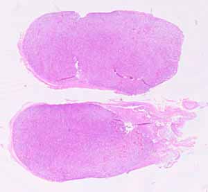
This may be one of the more difficult specimens in the SAQ series. At first glance, it may look like a complete jumble of cells with no apparent organization. Nevertheless (as with most specimens), there is no other organ quite like this one.
The slide carries sections from two similar pieces of the same organ.
Note the following:
- There is fibrous connective tissue enclosing the parenchyma. The entire organ is quite small, completely contained within this connective tissue.
- The organ is high vascular, with very many small vessels.
- The parenchymal cells are not uniform. Most have round or oval euchromatic nuclei, more-or-less centrally located. But the cytoplasm varies considerably. Some cells are quite acidophilic, others are more basophilic, still others stain less intensely.
- The parenchymal cells tend to occur in small irregular clusters, separated by thin stroma of connective tissue fibers and small vessels.
Hints on the next page are quite revealing. Don't look until you are ready to confirm your identification.
