
Olfactory epithelium
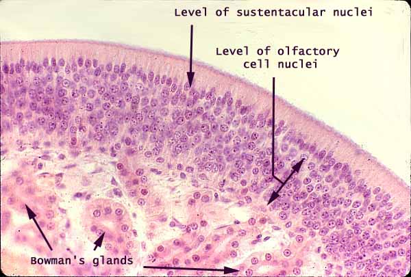
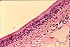
Olfactory epithelium cannot be properly appreciated in routine histological preparations. Even so, this pseudostratified, columnar epithelium can be readily distinguished from the respiratory epithelium which lines most of the airways.
Olfactory epithelium is thicker, with many more nuclei, than epithelium elsewhere in the respiratory tract. The only "proper" epithelial cells here are the "sustentacular" (sustaining or support) cells whose oval nuclei form the upper, outermost nuclear layer of this epithelium. Most of the remaining cells within the olfactory epithelium -- all those whose round nuclei lie deep to the outermost nuclear layer -- are olfactory receptor cells. These receptor cells share attributes of both epithelial cells and nerve cells. They have cilia at their apical ends, which are bathed in mucus; they also have axons extending from their basal ends. These axons pass through through the ethmoid bone's cribriform plate to form synapses in the olfactory lobe of the brain.
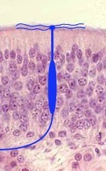
A cartoon of the shape of an olfactory cell is drawn in blue in the smaller image above. This shape, with its bizarre apical knob (olfactory vesicle) sprouting numerous, non-motile ciliary tentacles, is not apparent in routine micrographs, nor is the axon which proceeds from each olfactory cell to the olfactory bulb.
Mucus-secreting Bowman's glands are named after William Bowman (b. 1816, the same Bowman who gives his name to Bowman's capsules in the kidney).
Comments and questions: dgking@siu.edu
SIUC / School
of Medicine / Anatomy / David
King
https://histology.siu.edu/crr/CR004b.htm
Last updated: 1 May 2026 / dgk