Over subsequent decades, Bichat's nomenclature morphed into our familiar
set of four basic tissue types [c.f., Virchow],
each with several subdivisions.
Bichat did not trust microscopes, and hence did not practice "histology" in our modern "microscopic anatomy" sense. Although
microscopes had been used to good effect for over a century (cf., Hooke and Malpighi),
techniques for histological preparation were still rudimentary. Nevertheless, by subjecting tissues "successively to desiccation,
putrefaction, maceration, ebullition, stewing, and to the action of the acids and the alkalis" tissues could be separated "where the
scalpel was insufficient" [ 2 ]. Bichat might soak tissues for several months, or even swallow them
to be digested and subsequently regurgitated [ 2 ]. Thus was Bichat able to recognize that
organs were composed of more-fundamental simple materials, and furthermore to appreciate that pathology was commonly localized to or limited by specific types of tissue.
We conclude with two brief quotes. The first of these is a lightly edited excerpt from a note about a postage stamp.
The second is from the 1911 Encyclopedia Britannica, available
here, at Wikisource.
Baltic German anatomist, commemorated in "Boettcher's cells," among the many eponymous details associated with the
organ of Corti in the inner ear.
Boettcher (or Böttcher) was native to a town in the Russian empire which is now located in Latvia. He spent much of his
professional
career as professor of general pathology and pathological anatomy in Dorpat, in modern-day Estonia.
Curiously, reports by Boettcher in 1876 and 1877 appear to have introduced
a misconception which has endured for a century and a half, that camel red
blood cells have nuclei (unlike those of all other mammals). Even though reported as erroneous as long ago as
1928, this misconception persists and can still be easily found on the internet;
it has even appeared at least as recently as 2005 in the
prominent textbook Mammalogy. Boettcher's account of his studies of camel red blood cells can be found
in Mémoires de l'Académie Impériale de St.-Pétersbourg, VII Série. Tome XXII, No. 11 (1876), "Neue Untersuchungen über die rothen Blutkörperchen" and in
Archiv für mikroskopische Anatomie, xiv 73-93 (1877),
"Ueber die feineren Structurverhältnisse der rothen Blutkörperchen."
Bowman trained as a surgeon at Birmingham General Hospital
and at Kings College London, where he served as a prosector (preparing anatomical demonstrations). He was elected Fellow of the
Royal Society of London in 1841 for his work describing microscopical observations of muscle in a wide variety of species
[1]:
In 1842 Bowman presented to the Royal Society detailed microscopic
observations of the kidney in a variety of vertebrate animals: "On the structure and use of the Malpighian
bodies of the kidney" [2]. Bowman's
"Malpighian bodies" (i.e. "Malpighian corpuscles," first described by Marcello Malpighi two centuries
earlier) are now more commonly called renal corpuscles, containing
renal glomeruli. Malpighi had guessed, but could not
prove, that these bodies were connected with the tubules. Bowman was able to demonstrate that the eponymous epithelial capsule
around each renal glomerulus is the beginning of (i.e., continuous with) tubule epithelium, while the eponymous space is
continuous with the lumen of the associated tubule.
Bowman's contributions to understanding renal organization were so substantial, and his esteem
among his colleagues so high, that he was dubbed "the Father of the Kidney" [3]. However, Bowman misconstrued
the related functions of glomerulus and tubule:
In the same year (1842), an alternative [and more nearly correct] hypothesis was proposed by Bowman's contemporary
Carl Ludwig (German pioneer in physiology, 1816-1895), that renal glomeruli produce a
cell-free, protein-free filtrate of blood which is subsequently concentrated by renal tubules. Bowman's drawings of renal tubules were also
incomplete. They show proximal convoluted tubules leaving Bowman's capsule, but the loop of Henle is
missing. Description of that long tubular excursion into the renal medulla awaited the work of Jakob Henle a few
years later.
Bowman eventually specialized in and practiced ophthalmology, producing detailed, clinically oriented anatomical and histological
studies of the eye. His illustrations of the corneal limbus and the retina, published in "Lectures on the parts concerned in the operations
on the eye, and on the structure of the retina" [5], are more modern in appearance than his drawings of kidney.
During his career, Bowman corresponded with several eminent persons, including Florence Nightingale and Charles Darwin [6].
Darwin mentions his indebtedness to Bowman regarding the causes of weeping, in his The Expression of the Emotions in Man and Animals.
During Brodmann's relatively short professional career (he lived only 50 years) he was associated with several German universities
and other institutions. At the Municipal Mental Asylum in Frankfurt, he was influenced by A. Alzheimer (commemorated in
Altzheimer's disease) to pursue basic research in neuroanatomy.
In this work Brodmann surveyed the entire cortex, cataloging regional variations in the "cytoarchitecture" (detailed histological
appearance) of cortical layers. Brodmann's descriptions formed the original basis for recognizing what are now known as
Brodmann's areas. Significantly, Brodmann's areas are now known
to correspond with functional localization in the cortex (as presaged by the earlier work of Vladimir Betz).
Many resources for Brodmann, and for Brodmann's areas, are readily accessible on the internet. One especially satisfactory,
well-illustrated example, to which the interested reader is referred, is:
Bruch earned his medical doctorate at the University of Giessen in 1842 with a dissertation on granular pigments of animals,
which included his report of the eponymous membrane in the eye. He received an appointment to Heidelberg University in 1844,
where he assisted in the teaching of that institution's evolving histology courses as well as other anatomical disciplines.
To earn his habilitation for a professorship at Heidelberg, the university required that he translate his doctoral
dissertation into Latin. (Habilitation is a level of education above that of doctor, requiring the approximate
equilivent of a post-doctoral dissertion.) Bruch chose instead to write another dissertation, this one on rigor mortis.
Brunner was a student of Johann Jacob Wepfer (1620-1695), founder of the "Schaffhouse School" of anatomy and physiology,
in Schaffhausen, Switzerland. He worked with Wepfer and with Johann Conrad Peyer (discoverer in
1677 of his eponymous lymphoid follicles). In addition to describing glands of the duodenum and the pituitary gland,
in 1683 Brunner reported symptoms of diabetes experimentally induced by surgical removal of the pancreas in dogs.
But Brunner failed to associate his observations with the disease diabetes mellitus; making that connection remained for
physiologists von Mering and Minkowski two centuries later, in 1889.
Spanish neuroanatomist, professor at the University of Valencia, also of Barcelona and of Madrid; 1906 recipient of the
Nobel Prize in Physiology or Medicine.
Cajal is the most famous pioneer in the descriptive anatomy of nerve cells.
His elegant and precisely detailed drawings of individual neurons are quite familiar from their
appearance in many textbooks (e.g., the spectacular cerebellar Purkinje cell at right).
Cajal is commemorated in horizontal cells of Cajal in the cerebral cortex.
During the decades of Cajal's career, controversy raged over the nature of nervous tissue. Some, including Camillo Golgi, believed that
"ganglionic corpuscles" (now known as nerve cell bodies) were interconnected with one another through an
anastomosing reticulum of fibers. Others, including Cajal, became convinced by evidence
from many sources (see e.g., Cajal's
1906 Nobel Prize lecture) that each nerve cell body, together with its dendrites and axon, formed
a discrete cellular unit -- a "neuron" -- that was distinct and separate from other nerve cells.
Cajal's work elucidating nervous tissue came several decades after the cellular composition of most other tissues had been
fairly well understood. One simple reason for this is that no nerve cell type can be properly visualized in its
entirety in routine histological preparations.
Cajal was able to use the Golgi stain, a technique developed by his colleague and intellectual rival,
Camillo Golgi, to observe the shapes of individual nerve cells and infer not only their individuality but also the
direction of communication between them. Golgi, on the other hand, persisted in his conviction that nerve cells were not distinct
individuals but formed a reticulum. Both Cajal and Golgi shared the 1906 Nobel Prize for their work elucidating
nervous tissue. Their conflicting views are exhibited in their respective Nobel Prize acceptance speeches:
Cajal /
Golgi.
Cajal introduced the four
principles which comprise the neuron doctrine (quotations here are taken from Eric Kandel's 2006 autobiography
In Search of Memory, pp. 65-66;
full text here):
- Cellularity: "The nerve cell is the fundamental
structural and functional element of the brain."
- Synaptic communication: "The terminals of
one neuron's axon communicate with the dendrites of another neuron only
at specialized sites, later named synapses by
Sherrington."
- Connection specificity: "Neurons do not
form connections indiscriminately. Rather each nerve cell forms
synapses and communicates with certain nerve cells and not with others."
- Dynamic polarization: "Signals in a neural
circuit travel in only one direction. . . Information flows, from
the dendrites of a given nerve cell to the cell body [then] along the
axon to the presynaptic terminals and then across the synaptic cleft to
the dendrites of the next cell, and so on."
Cajal was profoundly impressed by the beauty and diversity of nerve cells as revealed by Golgi impregnation, referring to them
in his autobiography as "the mysterious butterflies of the soul" ["las misteriosas mariposas del alma"]. Regarding fame, he once wrote, "And what do
praises matter to me? When they applaud me I will not exist" [quote taken from Gomez-Marin's review of Benjamin Ehrlich's 2022 biography, cited below].
Sources for images by Cajal:
Additional resources:
- A recent eloquent biography in print: The Brain in Search of Itself: Santiago Ramón y Cajal and the Story of the Neuron, by Benjamin Ehrlich;
Farrar, Straus and Giroux, 2022. For a sample of Ehrlich's writing about Cajal, see The Paris Review.
This biography is reviewed in Science, 375: 1237 (March 18, 2022), "Drawing the mind,
one neuron at a time," by Alex Gomez-Marin.
- Biographical sketch of Cajal, from the Nobel Prize Organization.
- Biography of Cajal, from Wikipedia.
- Golgi's method, used by Cajal, from Wikipedia.
- Cajal's 1917 autobiography, Recollections of My Life [Recuerdos de mi Vida].
Volume 1 (text in Spanish), from Project Gutenberg.
Volume 2 (text in Spanish), from Project Gutenberg.
Review of 1937 English translation, in Nature.
- Foundations of the Neuron Doctrine: 25th Anniversary Edition, 2nd Edition, by Gordon M Shepherd. Oxford Univ. Press 2015.
[preview]
A B C D
E F G H
I J K L
M N O P
Q R S T
U V W X
Y Z
TOP OF PAGE / ALPHABETICAL INDEX / CHRONOLOGICAL INDEX
Max Clara (1899-1966)
German doctor whose research exploited doomed prisoners of the Nazi regime prior
to and during WWII. Older textbooks refer to bronchiolar exocrine cells with his eponym, but
because of this tainted history the alternative term club cells
has been adopted by several journals and societies.
Issues associated with morally-questionable eponymy are addressed in an essay by Rachel E. Gross, "Should Medicine Still Bother
With Eponyms?" (The New York Times, 19 June 2023),
which asks whether expunging eponyms commemorating Nazi-era doctors will lead physicians to forget lessons of the past: "some scholars
contend that even 'canceled' eponyms have a place, as stark reminders of the ethical breaches medicine should never repeat."
A similar ethical issue has arisen in the fields of zoological and botanical nomenclature:
Should beetles be named after Adolf Hitler? (Science, doi: 10.1126/science.adk6936)
Additional resources:
H. Czech et al. (2021) From scientific exploitation to individual memorialization: Evolving attitudes towards research on Nazi victims' bodies. Bioethics 35(6):508-517. doi: 10.1111/bioe.12860.
A. Winkelmann, T. Noack (2010) The Clara cell: a "Third Reich eponym"? Respir. J. 36(4):722-7. doi: 10.1183/09031936.00146609. .
Hildebrandt, Sabine, The Anatomy of Murder: Ethical Transgressions and Anatomical Science during the Third Reich, Berghahn Books, 2016.
Biography with additional references at Wikipedia.
A B C D
E F G H
I J K L
M N O P
Q R S T
U V W X
Y Z
TOP OF PAGE / ALPHABETICAL INDEX / CHRONOLOGICAL INDEX
Friedrich Matthias Claudius (1822-1869)
German zoologist and anatomist, commemorated by "cells of Claudius" associated with the organ of Corti in the inner ear.
Claudius held positions at the Zoological Museum of Kiel University and at the anatomical
institute at the University of Marburg.
(For more on cells of Claudius, as well as on other eponymous inner-ear structures, see
J. Schacht & J.E. Hawkins,
"A Cell by Any Other Name: Cochlear Eponyms," Audiology & Neuro Otology 2004, vol. 9, pp. 317-327, DOI: 10.1159/000081311.)
Very brief bio at Wikipedia.
Very brief bio at Cochlear Explorers.
(This bio ends with, "Not much else is known about the person who first described the Cells of Claudius.")
Publications by Claudius
- 1856: Bemerkungen über den Bau
der häutigen Spiralleiste der Schnecke ["Remarks on the structure of the spiral strip of the cochlea"], Zeitschrift für wissenschaftliche Zoologie, vol. 7, pp. 154-161 (this
manuscript reports the eponymous cells).
- 1867:
Das Gehörorgan von Rhytina stelleri ["The hearing organ of Steller's sea cow"].
A B C D
E F G H
I J K L
M N O P
Q R S T
U V W X
Y Z
TOP OF PAGE / ALPHABETICAL INDEX / CHRONOLOGICAL INDEX
Alfonso Giacomo Gaspare Corti (1822-1876)
Italian nobleman-physician who worked in Germany and published in French. Corti is commemorated in the organ of
Corti within the cochlea of the inner ear.
Corti published his "pivotal paper" describing the eponymous organ in 1851, while he was working in the laboratory
of "the father of modern histology," Albert von Kölliker. Corti "retired
from scientific investigation the year after his reporting the description of the organ of Corti to assume his
new role as Baron Corti following the death of his father" [1]. Corti's description
"was soon followed by papers on the descriptive anatomy of the cochlear receptor by Professors Claudius (1856),
Deiters (1860), Hensen (1863),
Boettcher (1869), and Nuel (1872)" [1] (each
of whom has his own eponymous part of the cochlea); in 1863 Kölliker himself described the eponymous
"Kölliker's organ," the embryonic precursor of epithelial structures in the organ of
Corti.
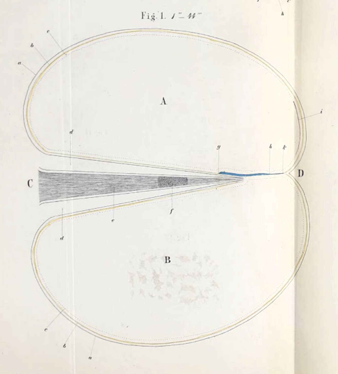

Corti's histological protocols were inadequate for preserving and displaying all the associated cells and structures of
the cochlea; in particular Reissner's membrane is missing from these illustrations. Consequently, his diagrammatic
illustrations [2] are somewhat difficult to interpret; in the two
images reproduced here, the spiral lamina is on the left and the basilar membrane is on
the right. But subsequent researchers were able to add more and more detail (which resulted in more and more eponyms).
[1] Quotes above are from p. 1743 in: van de Water,
Historical aspects of inner ear anatomy and biology, The Anatomical Record, vol. 295, pp. 1741-1759 (2012). This paper is
also a rich source for inner-ear anatomy and physiology.
[2] Images above are from: Corti A. (1851), "Recherches sur l'organe de l'ouïe des mammiféres"
[Research on the organ of hearing of mammals], Zeitschrift für wissenschaftliche
Zoologie 3:109-169, available at the Wellcome Collection.
Brief biography at Wikipedia.
The following articles are available only to subscribers to the journals:
(For more on the organ of Corti, as well as on other eponymous inner ear structures, see
J. Schacht & J.E. Hawkins,
"A Cell by Any Other Name: Cochlear Eponyms," Audiology & Neuro Otology 2004, vol. 9, pp. 317-327, DOI: 10.1159/000081311.)
A B C D
E F G H
I J K L
M N O P
Q R S T
U V W X
Y Z
TOP OF PAGE / ALPHABETICAL INDEX / CHRONOLOGICAL INDEX
William Cowper (1666-1709)
English anatomist, commemorated in Cowper's glands (bulbourethral glands) of
the male reproductive tract.
Cowper is noted for an elegantly illustrated anatomy atlas, whose full title page,
somewhat obscured by multiple fonts
in the image at right, reads:
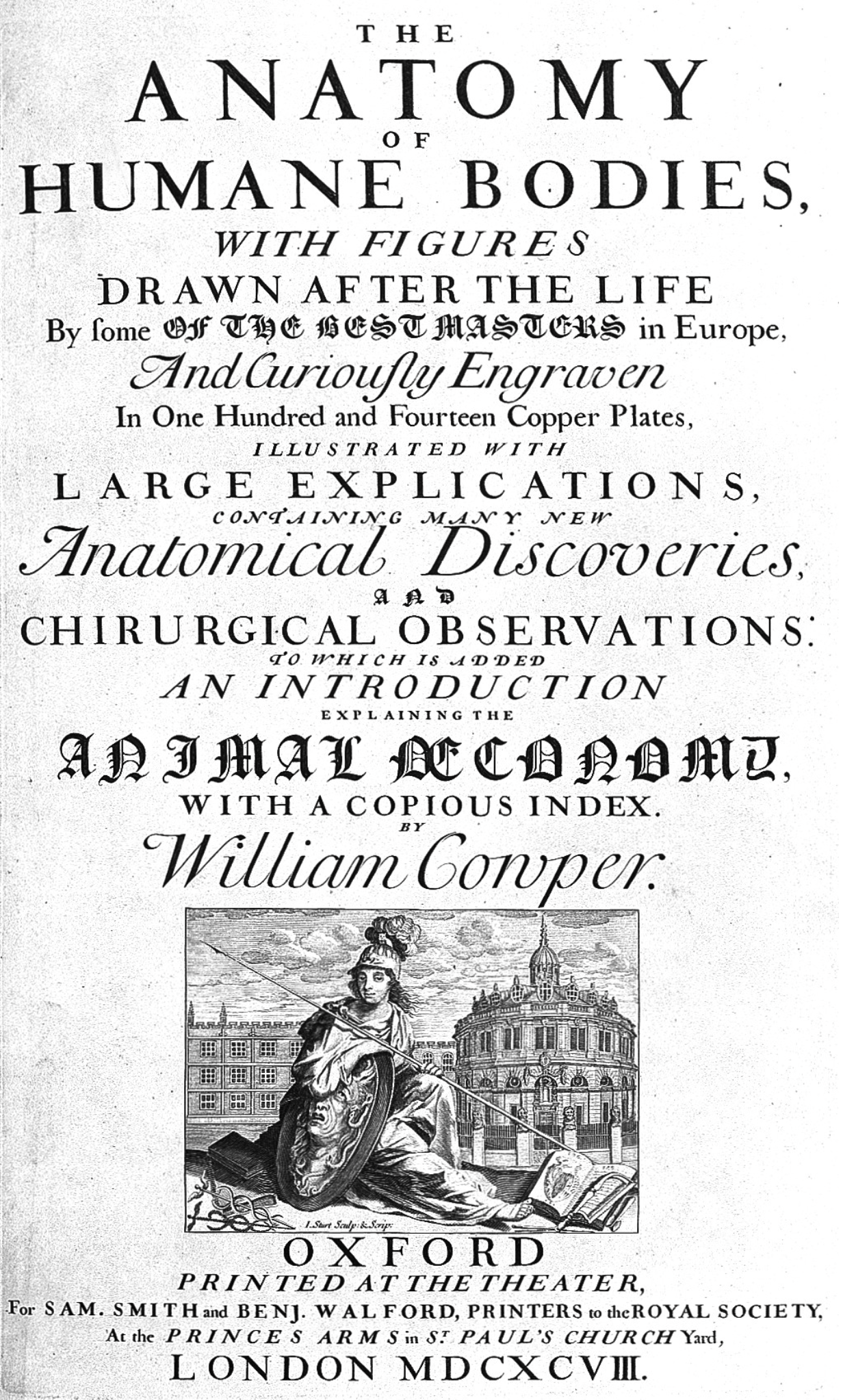
THE
A N A T O M Y
OF
HUMANE BODIES,
WITH FIGURES
DRAWN AFTER THE LIFE
By some OF THE BEST MASTERS in Europe,
And Curiously Engraven
In One Hundred and Fourteen Copper Plates,
ILLUSTRATED WITH
LARGE EXPLICATIONS,
CONTAINING MANY NEW
Anatomical Discoveries,
AND
CHIRURGICAL OBSERVATIONS:
TO WHICH IS ADDED
AN INTRODUCTION
EXPLAINING THE
ANIMAL ŒCONOMY,
WITH A COPIOUS INDEX.
by
William Cowper.
In this title, Chirurgical = "surgical." A digital facsimile of this volume is available at the
Internet Archive.
Cowper's atlas is notorious as "one of the greatest acts of plagiarism in medical publishing history." Although the text
of this atlas did contribute substantially to the field of anatomy, the plates had been purchased from the publisher of other anatomists'
works. Cowper's failure to give adequate credit for the engravings created a scandal. That scandal in turn contributed
to the evolution of copyright law. This story is told in greater detail in the resources below.
Digital
exhibit on Cowper and his Anatomy, at the University of Guelph.
Brief essay about Cowper's Anatomy, with sample illustrations, at Saint Louis
University Special Collections.
Older, and lengthier,
1887 essay at The Dictionary of National Biography.
Brief biography at Wikipedia.
A B C D
E F G H
I J K L
M N O P
Q R S T
U V W X
Y Z
TOP OF PAGE / ALPHABETICAL INDEX / CHRONOLOGICAL INDEX
 Otto Deiters (1834-1863)
German neuroanatomist, commemorated in Deiters' nucleus (the lateral vestibular nucleus) of the brainstem and Deiters' cells in the organ
of Corti of the inner ear.
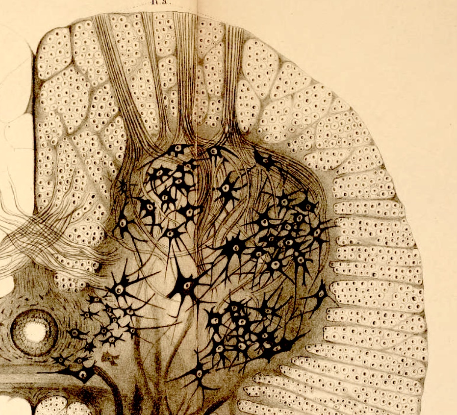
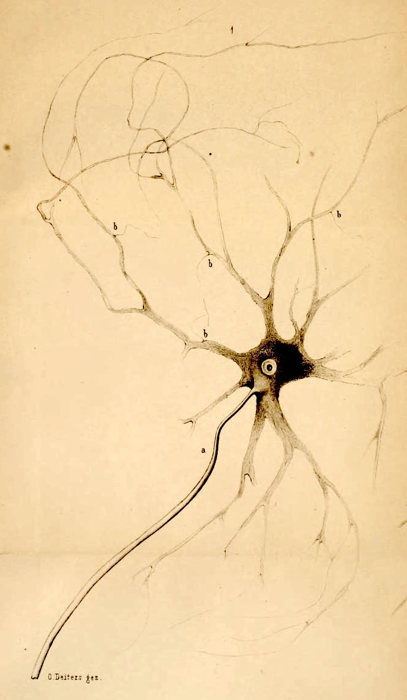
During his short life (he died of typhoid at age 29) Deiters produced a thick monograph on his research into the brain and spinal cord of humans and mammals
(Untersuchungen über Gehirn und Rückenmark des Menschen und der Säugethiere, 1865), from which the illustrations here were taken.
The image at left illustrates the anterior horn of the spinal cord (anterior is up).
Note in white matter the stylized representation of myelinated axons
as black dots (stained) surrounded by clear halos (unstained). The image at right shows
a spinal motor neuron, with the axon emphasized.
Working as he did before establishment of the Neuron Doctrine, Deiters' interpretations
were limited by understanding of his time. Nevertheless he produced wonderfully detailed images (as shown here) that
display how far neurohistology had progressed prior to the work of Ramón y Cajal.
(For further discussion of the state of neurohistological knowledge during the nineteenth century,
see "The Discovery of the Neuron," by Mo
Costandi, at the Neurophilosophy Blog.)
Within the inner ear, Deiters' cells are supporting cells in the organ of
Corti. Deiters' cells are situated between hair cells and the basilar membrane.
For more on Deiters' cells, as well as on other eponymous inner ear structures, see:
Publications by Deiters
- Untersuchungen über die Lamina spiralis membranacea: ein Beitrag zur Kenntniss des inneren Gehörorgans [Studies on the lamina spiralis membranacea: a contribution to knowledge of the inner ear] (1860):
Available from the Wellcome Collection.
Available from the Internet Archive.
- Untersuchungen über Gehirn und Rückenmark des Menschen und der Säugethiere [Studies on the brain and spinal
cord of man and mammals] (1865), edited after Deiters' death by Max Schultze:
Available from the Internet Archive.
Available from the Wellcome Collection
Brief biography at Wikipedia
A B C D
E F G H
I J K L
M N O P
Q R S T
U V W X
Y Z
TOP OF PAGE / ALPHABETICAL INDEX / CHRONOLOGICAL INDEX
Jean Descemet (1732-1810)
French physician, commemorated in Descemet's membrane of
the cornea, which he described in 1758 in his graduate thesis on the anatomy of the cornea and lens,
submitted for his doctorate.
At about the same time (1759), pursuing his interest in botany,
Descemet published Catalogue des plantes du jardin de MM. les apothicaires de Paris, suivant leurs genres
et les caractères des fleurs, conformément à la méthode de M. Tournefort
[Catalog of the plants in the Garden of the Apothecaries in Paris according to their genera and the characteristics of their
flowers, using Mr. Tournefort's method].
Wikipedia offers only a very brief biographical entry, here.
A more extensive account is available in the "Descemet" entry in
The Dawn of Modern Medicine: An Account of the Revival of the Science and Art of Medicine Which Took Place in Western Europe
During the Latter Half of the Eighteenth Century and the First Part of the Nineteenth,
by Albert Henry Buck (Yale Univ. Press, 1920), accessed at Project Gutenberg.
A B C D
E F G H
I J K L
M N O P
Q R S T
U V W X
Y Z
TOP OF PAGE / ALPHABETICAL INDEX / CHRONOLOGICAL INDEX
Joseph Hugo Vincent Disse (1852-1912)
German experimental pathologist who demonstrated in the liver the existence of a space filled
with lymph (= blood plasma) between blood-filled hepatic sinusoids and their underlying hepatocytes,
the eponymous space of Disse.
Because Disse worked long before electron microscopy became a practical tool for histology
(beginning ca. 1939), he was not able to visually resolve the endothelium with its fenestrations.
Nevertheless his experiments convincingly established the presence of a functional barrier at the location
of the sinusoidal endothelium.
[More
: A fascinating description of Disse's demonstration of the functional distinction between
liver sinusoids and his eponymous space is presented in a short biographical essay,
"Who Was
Disse," by Rudi Schmid, in the journal Hepatology, vol. 14, pp. 1283-1285, 1991.]
A B C D
E F G H
I J K L
M N O P
Q R S T
U V W X
Y Z
TOP OF PAGE / ALPHABETICAL INDEX / CHRONOLOGICAL INDEX
Sigmund Freud (1856-1939)
Austrian neuroanatomist at the beginning of his professional career; later, of course, Freud became a famed psychoanalyst. Not commemorated in any histological eponyms.
I couldn't resist including Freud here. Before becoming a world-famous psychiatrist, Freud began his career with pioneering histological studies on the
neuronal cytoskeleton in axons of crayfish. I first discovered Freud's research on nerve cells
while preparing my own doctoral dissertation (1975) on the organization of crustacean neuropil.
Great
moments in crayfish research: Before he was famous, by
Zen Faulkes
Sigmund
Freud's contribution to the history of the neuronal cytoskeleton, by E. Frixione,
J. Hist Neurosci. 2003, 12:12-24. doi: 10.1076/jhin.12.1.12.13790.
The Cytoskeleton
of Nerve Cells in Historical Perspective, by E. Frixione, IBRO History of Neuroscience, 2006.
Freud, Sigmund (1882) Über den Bau der Nervenfasern und Nervenzellen
beim Flusskrebs.
Sitzungsberichte der kaiserliche Akademie der Wissenschaften (Wien)
85: 9-46.
For Freud's taste in microscopes, see http://www.microscopy-uk.org.uk.]
Wikipedia's very extensive entry on Freud includes only one sentence that
mentions crayfish: "In 1877, Freud moved to Ernst Brücke's physiology laboratory where he spent six years comparing the brains
of humans and other vertebrates with those of frogs and invertebrates such as crayfish and lampreys."
A B C D
E F G H
I J K L
M N O P
Q R S T
U V W X
Y Z
TOP OF PAGE / ALPHABETICAL INDEX / CHRONOLOGICAL INDEX
|
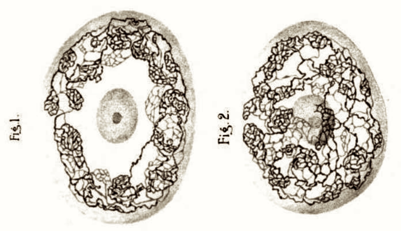
Golgi apparatus illustrated in a dorsal root ganglion neuronal cell body
(from p. 674 in Golgi's 1903 Opera Omnia, Vol. 2, accessed at GoogleBooks).
|
|
|
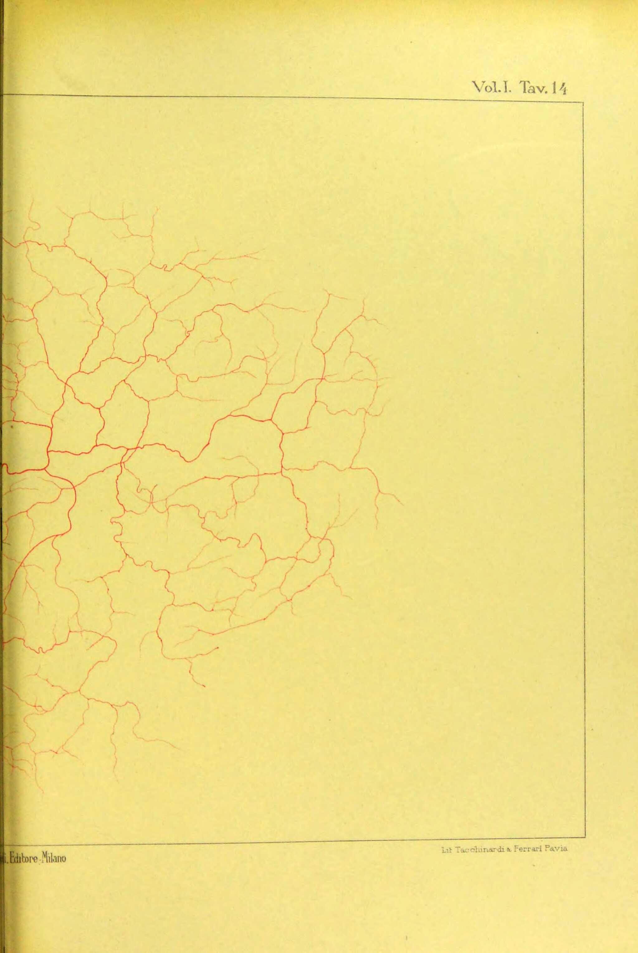
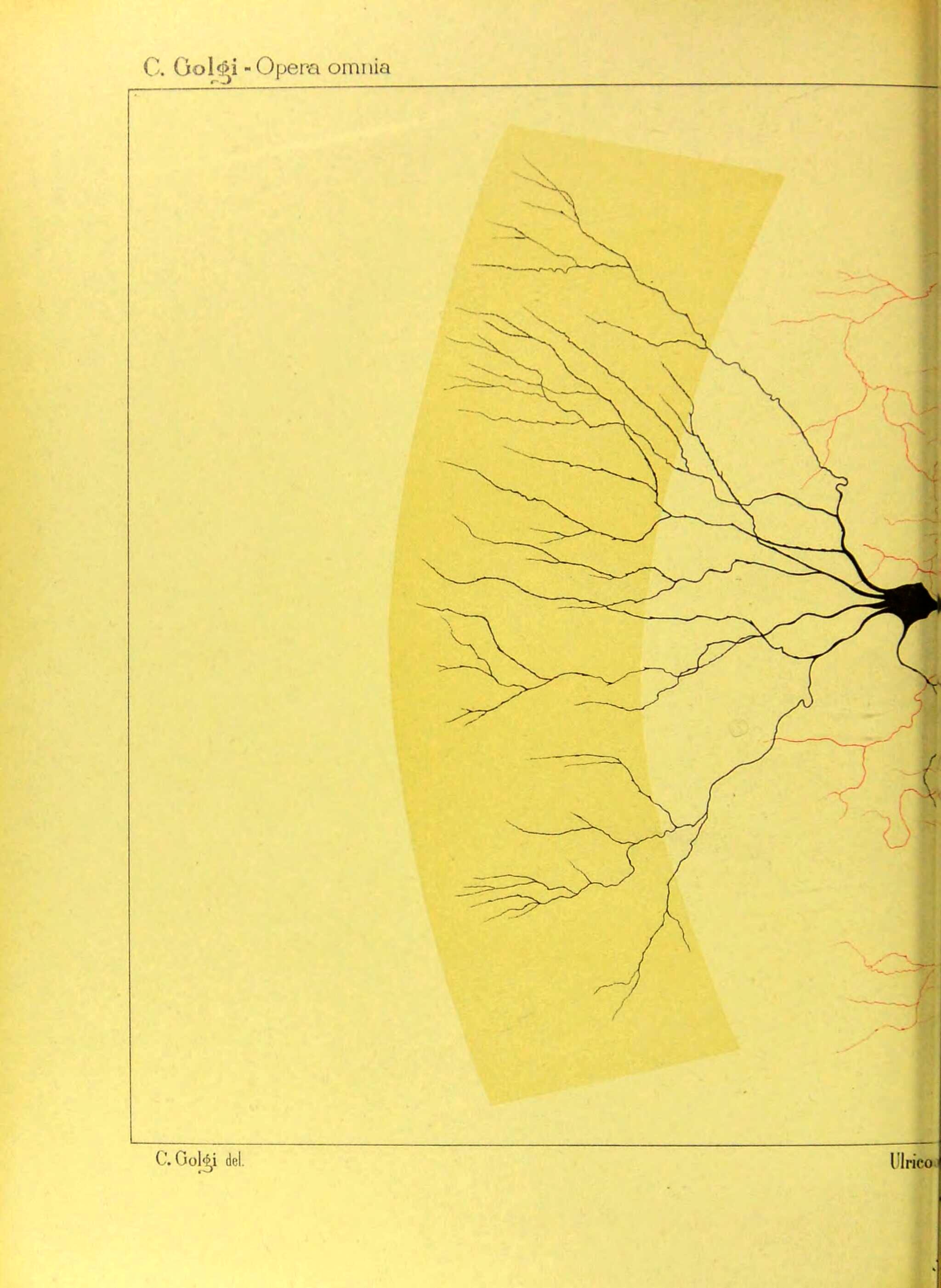
These images (from pp. 382 and 383 in Golgi's 1903 Opera Omnia, Vol. 1, accessed at The Wellcome Collection) show one of the cerebellar neurons of a type
now called "Golgi cells."
|

|
|
Golgi tendon organ (p. 205 from Golgi's
1903 Opera Omnia, accessed at The Wellcome Collection).
|
Camillo Golgi (1843-1926)
Italian pathologist and histologist, commemorated in several eponyms, most notably the Golgi stain
and the Golgi apparatus; also Golgi type 1 (long axon) neurons, Golgi type 2 (short axon) neurons, Golgi tendon organs, and Golgi cells of cerebellum.
Additional Golgi eponyms can be found at
Whonamedit.com.
Golgi's most notable contribution to histology was the discovery not of a structure but of a technique, la reazione nera
("the black reaction"), which used potassium dichromate and silver nitrate to produce a black precipitate within particular
structures. The deposition of precipitate is quite variable and difficult to control, but in skilled hands results can be
powerfully revealing. Golgi's black reaction became famous as "Golgi impregnation" or "Golgi staining."
For more about Golgi staining, see
Wikipedia.
With this stain, Golgi discovered his apparato reticolare interno ("internal reticular apparatus"). This structure,
which is especially elaborate in nerve cell bodies (e.g., top illustration at right), has become known as the
Golgi apparatus. Because results from the Golgi stain can be erratic, this structure remained suspect as a possible
artifact until its reality was convincingly demonstrated decades later, by electron microscopy. The Golgi apparatus is now
understood to be an essential component of most cells, so that Golgi's name has become an integral part of the vocabulary of cell biology.
For more about the Golgi apparatus, see
www.genome.gov (brief definition),
Wikipedia (extensive account), or
National Library of Medicine (cell biology textbook chapter).
Golgi applied his stain to extensive pioneering studies of nervous tissue, as did his contemporary and intellectual rival
Santiago Ramón y Cajal. Both men were honored together by the 1906 Nobel Prize in Physiology or
Medicine, "in recognition of their work on the structure of the nervous system." Golgi's reputation in neuroscience was
subsequently eclipsed by that of Cajal, primarily because Golgi stood on the wrong side of history in his understanding of neural
organization. Even as evidence to the contrary was accumulating, Golgi persisted in his belief that nervous tissue was a syncytium,
an anastomosing reticulum with cell bodies sharing cytoplasmic connections. (Golgi's syncytial understanding of nervous tissue was
revisited in 2023, when a report [Science 380:241--242] suggested that
a syncytial nerve net might actually exist in some ctenophores -- an animal group only distantly related to all other creatures.) Cajal,
in contrast, understood that nerve cells were distinct entities, each with long axonal and dendritic processes that made contact
with other nerve cells at synapses but without cytoplasmic continuity, an understanding that became known as the
"Neuron Doctrine."
These conflicting views are exhibited in their respective Nobel Prize acceptance speeches:
Golgi /
Cajal.
A tidy summary of this neuroscience story can be found here, in a
book review by Mitchell Glickstein of P. Mazzarello's 2009 biography, "Golgi: A Biography of the Founder of Modern Neuroscience"
(ISBN: 978-0-19-533784-6).
Additional resources:
- Golgi's collected works, published in Opera Omnia, 1903,
at the Wellcome Collection.
- Biographical entry from the Nobel Prize website.
- "Life and Discoveries of
Camillo Golgi," from the Nobel Prize website, including a few images from Opera Omnia.
- Extensive biographical entry at Wikipedia, including a brief account of
Golgi's work contributing to understanding of malaria.
- "The Centenarian Golgi apparatus,"
an account of discovery, with images by Golgi, from Nature, May 1998 DOI: 10.1038/33266.
- "1898: The Golgi apparatus emerges from nerve cells,"
from Trends in Neuroscience.
- "The Original
Histological Slides of Camillo Golgi and His Discoveries on Neuronal Structure," by M. Bentivoglio
et al., in Frontiers in Neuroanatomy, Vol. 13, article 3 (2019).
A B C D
E F G H
I J K L
M N O P
Q R S T
U V W X
Y Z
TOP OF PAGE / ALPHABETICAL INDEX / CHRONOLOGICAL INDEX
Norbert Goormaghtigh (1890-1960)
Belgian physiolologist, commemorated in Goormaghtigh cells
of the renal juxtaglomerular apparatus.
A detailed account of Goormaghtigh's investigations into the histophysiology of the juxtaglomerular apparatus (which he named)
can be found in "The juxtaglomerular
apparatus of Norbert Goormaghtigh -- a critical appraisal," by G. Eknoyan, et al. (2009) Nephrology
Dialysis Transplantation, V. 24, pp. 3876-3881 (https://doi.org/10.1093/ndt/gfp503).
Additional information:
"Norbert Goormaghtigh and his contribution to the
histophysiology of the kidney," by Hendrik Roels (2003), Journal of Nephrology, 16: 965-9.
Very brief biography at Wikipedia
(in Dutch, easily translated by copy-and-pasting into GoogleTranslate.
A B C D
E F G H
I J K L
M N O P
Q R S T
U V W X
Y Z
TOP OF PAGE / ALPHABETICAL INDEX / CHRONOLOGICAL INDEX
Reinier de Graaf (1641-1673)
Dutch physician and anatomist, commemorated in ovarian Graafian follicles.
De Graaf studied reproductive anatomy and physiology during a
century when biologists were only beginning to understand the basic principles of animal reproduction.
The eponymous follicles were initially named ova Graafiana by Albrecht von Haller, pioneering physiologist who believed the follicle itself was the ovum. (Spermatozoa were first reported by Leeuwenhoek
shortly after De Graaf's death. Karl Ernst von Baer is credited with finally
observing mammalian oocytes in 1827.)
Additional biographical information:
Reinier De Graaf (1641-1673) and the
Graafian follicle, by M. Thiery, Gynecological Surgery, vol. 6, pp. 189-191 (2009).
Brief biography, at The Embryo Project.
Brief biography, at Wikipedia
A B C D
E F G H
I J K L
M N O P
Q R S T
U V W X
Y Z
TOP OF PAGE / ALPHABETICAL INDEX / CHRONOLOGICAL INDEX
William Harvey (1578-1657)
English physician, in an era before microscopes; best known for his discovery* of the circulation of blood.
* In a letter to The New Yorker (24 March 2025, p. 9), a writer who happens to share my name (David King), pointed
out that Harvey's demonstration of blood circulation was preceded by a 1553 description of pulmonary circulation by Spanish physician,
Miguel Servet; Servet's work in turn might have been based on that of a
thirteenth-century Arab researcher, Ibn al-Nafis.
In this "histology eponyms" webpage, this is the only entry acknowledging the important historical contribution to medicine of Islamic
research. Islamic influence on the emergence of modern science had waned long before the invention of microscopes and
the advent of histology as a discipline.
Although no histological eponyms are associated with Harvey, his works include a list of tissue elements which loosely presages
the pioneering work of Bichat (the "Father of Histology). Bichat famously listed
21 simple tissue types. Harvey's list includes substances in four broad categories: "(a) liquids: blood, sperm, milk, ocular humours, rheum, bile, mucus, tears,
ichor, serum; (b) solids: (i) soft: flesh of muscle, of gums etc., of parenchyma, and of glands, marrow, fat, lard, brain, lens of eye;
(ii) firmer: fibre, membrane, vein, artery, skin, nerve, tendon, ligament;
(iii) hard: bone, teeth, carapace, hair, cartilage, nail, claw, horn, quill, beak, feathers, scales" (as quoted in
[ 1 ]).
Harvey served as personal physicial to King Charles I. In 1628 he presented his theory
of blood circulation in a letter addressed "To The Most Illustrious And Indomitable Prince Charles
King Of Great Britain, France, And Ireland Defender Of The Faith."
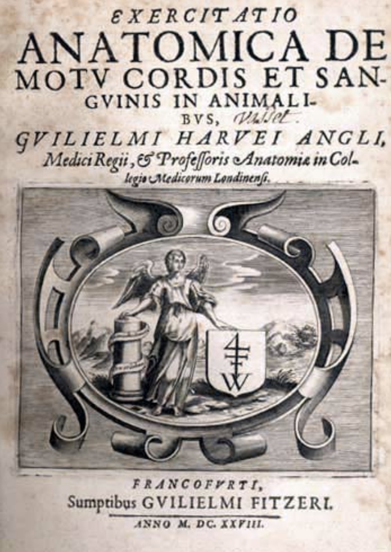
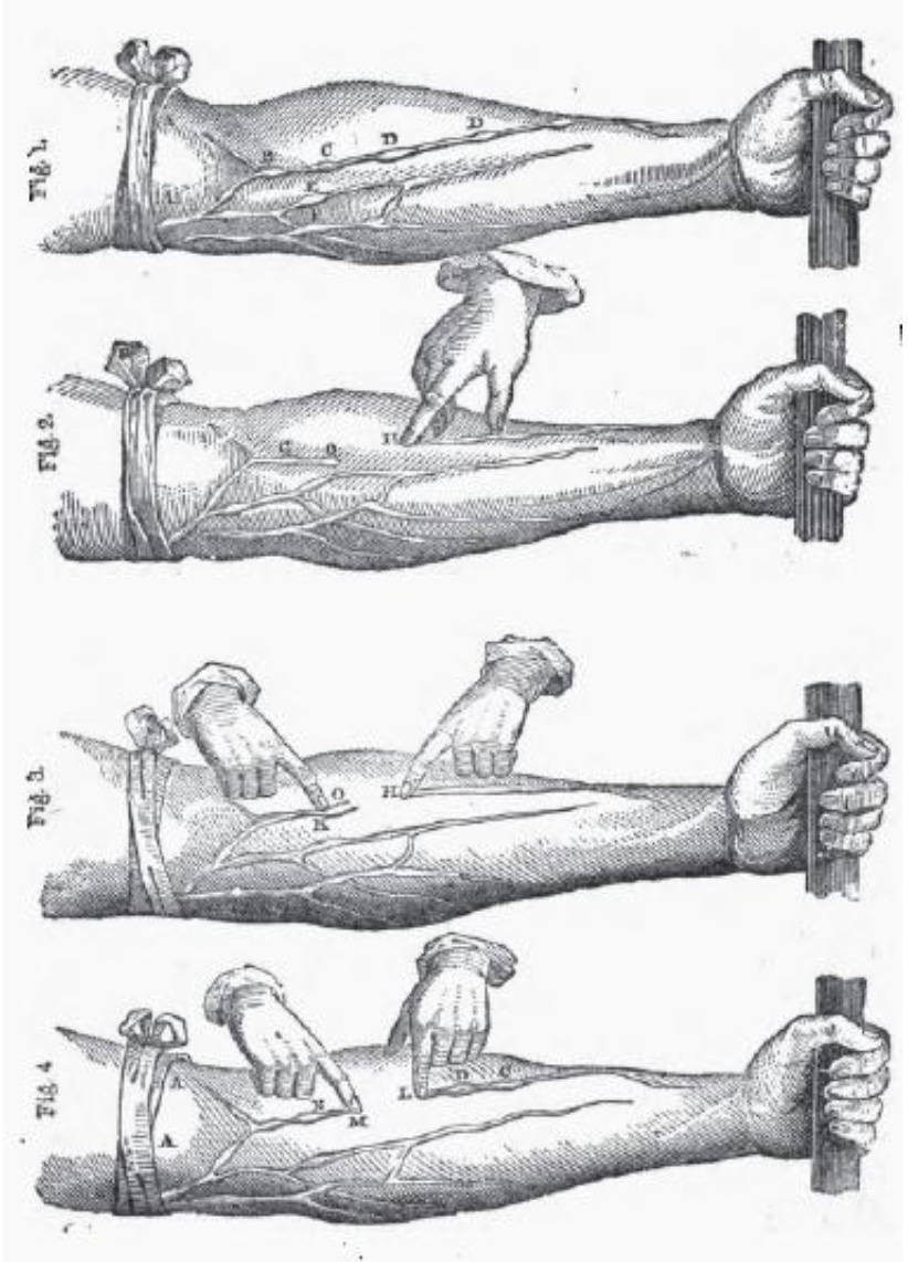 This letter, Exercitatio Anatomica de Motu Cordis et
Sanguinis in Animalibus [ 2 ], included a nice illustration
to guide easy classroom demonstration of the presence and function of valves in forearm veins:
"...[L]et an arm be tied up above the elbow as if for phlebotomy (A, A, fig. 1). At intervals in the course of
the veins ... certain knots or elevations (B, C, D, E, F) will be perceived... [T]hese knots or risings are all formed by valves, which
thus show themselves externally. And now if you press the blood from the space above one of the valves, from H to O, (fig. 2,)
and keep the point of a finger upon the vein inferiorly, you will see no influx of blood from above; the portion of the vein between
the point of the finger and the valve O will be obliterated; yet will the vessel continue sufficiently distended above the valve
(O, G). The blood being thus pressed out and the vein emptied, if you now apply a finger of the other hand upon the distended
part of the vein above the valve O, (fig. 3,) and press downwards, you will find that you cannot force the blood through or beyond the
valve; but the greater effort you use, you will only see the portion of vein that is between the finger and the valve become more
distended, that portion of the vein which is below the valve remaining all the while empty (H, O, fig. 3)." [ 2 ]
In spite of his friendship with King Charles, Harvey's understanding of blood circulation was not widely adopted during his lifetime.
A poignant account of Harvey's difficult professional career through the English Civil War, including his time as a Royalist during the siege
of Oxford, can be found in Soul Made Flesh, by Carl Zimmer [ 3 ].
Most significantly for the history of histology, Harvey predicted the necessity of invisibly small pores
(i.e., capillaries) as an essential corollary of his theory of blood
circulation. Capillaries were actually illustrated by Marcello Malpighi in 1661. Unfortunately Harvey,
who died in 1657, did not live quite long enough to see this confirmation of his theory.
Wikipedia offers a more extensive biography.
Also see "
Completing the puzzle of blood circulation:
the discovery of capillaries," from ResearchGate. For an extended account of subsequent history into the nature
of capillaries, see "The history of the capillary
wall: doctors, discoveries, and debates," by C. Hwa and W.C. Aird, in Am J Physiol Heart Circ Physiol 293: H2667-H2679 (2007),
doi:10.1152/ajpheart.00704.)
| Cited references: |
- Harvey's list of tissue elements, from The anatomical lectures of William Harvey, is quoted in footnote 50 on p. 450 of
Forrester, "The homoeomerous parts and their replacement by Bichat's tissues,"
Medical History, 1994, 38: 444-458.
- On The Motion Of The Heart And Blood In Animals, 1628;
translation from original Latin by Robert Willis, 1847 (from Fordham University).
- Soul Made Flesh, Carl Zimmer, 2004, pp. 64-76, 117-120.
|
A B C D
E F G H
I J K L
M N O P
Q R S T
U V W X
Y Z
TOP OF PAGE / ALPHABETICAL INDEX / CHRONOLOGICAL INDEX
Arthur Hill Hassall (1817-1894)
British physician (general practitioner) commemorated in Hassall's corpuscles of thymus.
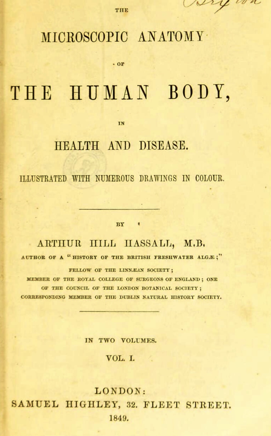
In 1849, Hassall published The microscopic anatomy of the human body, in health and disease, in two volumes, the first English-language textbook of microscopic anatomy.
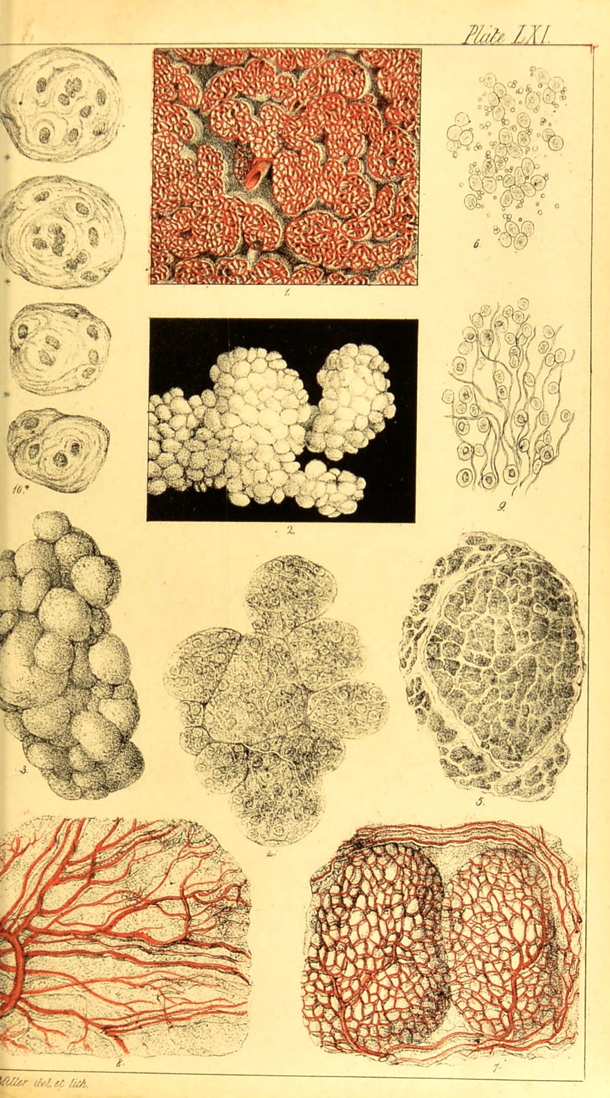 |
| Hassall's Plate LXI in Vol. 2 illustrates thymus; the small sketches in the upper left corner represent the
eponymous corpuscles, referred to by the author as "compound cells."
|
From Hassall's preface: "At the time when [the design of this
work] was first conceived, the powers of the microscope in developing organic structure were but beginning to be known and appreciated, and the
importance of its application to physiology and pathology was but dimly perceived... Now, one of the purposes [of this work] has been the collecting
together of the numerous communications on general anatomy to be found scattered through the pages of our different scientific periodicals and their
combination into a whole."
This textbook occupies a fascinating place in the history of histology, decades after Bichat's pioneering work (which
had been completed without use of microscope) but only a few years after Schwann's establishment of Cell Theory.
Thus this book represents knowledge of microscopic anatomy prior to the development of effective techniques for preparing, sectioning, and staining
tissue specimens.
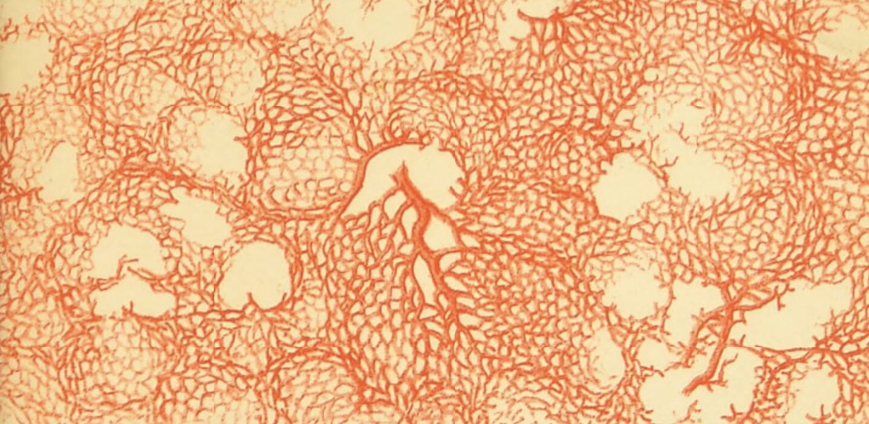 The numerous illustrations in Hassall's volumes are often based on views of fine dissections and vascular injections rather
than sections, and these commonly emphasize capillary beds. (Scanning Hassell's
INDEX OF THE ILLUSTRATIONS is a worthwhile exercise, to gain an appreciation for the incredibly comprehensive coverage of this work.)
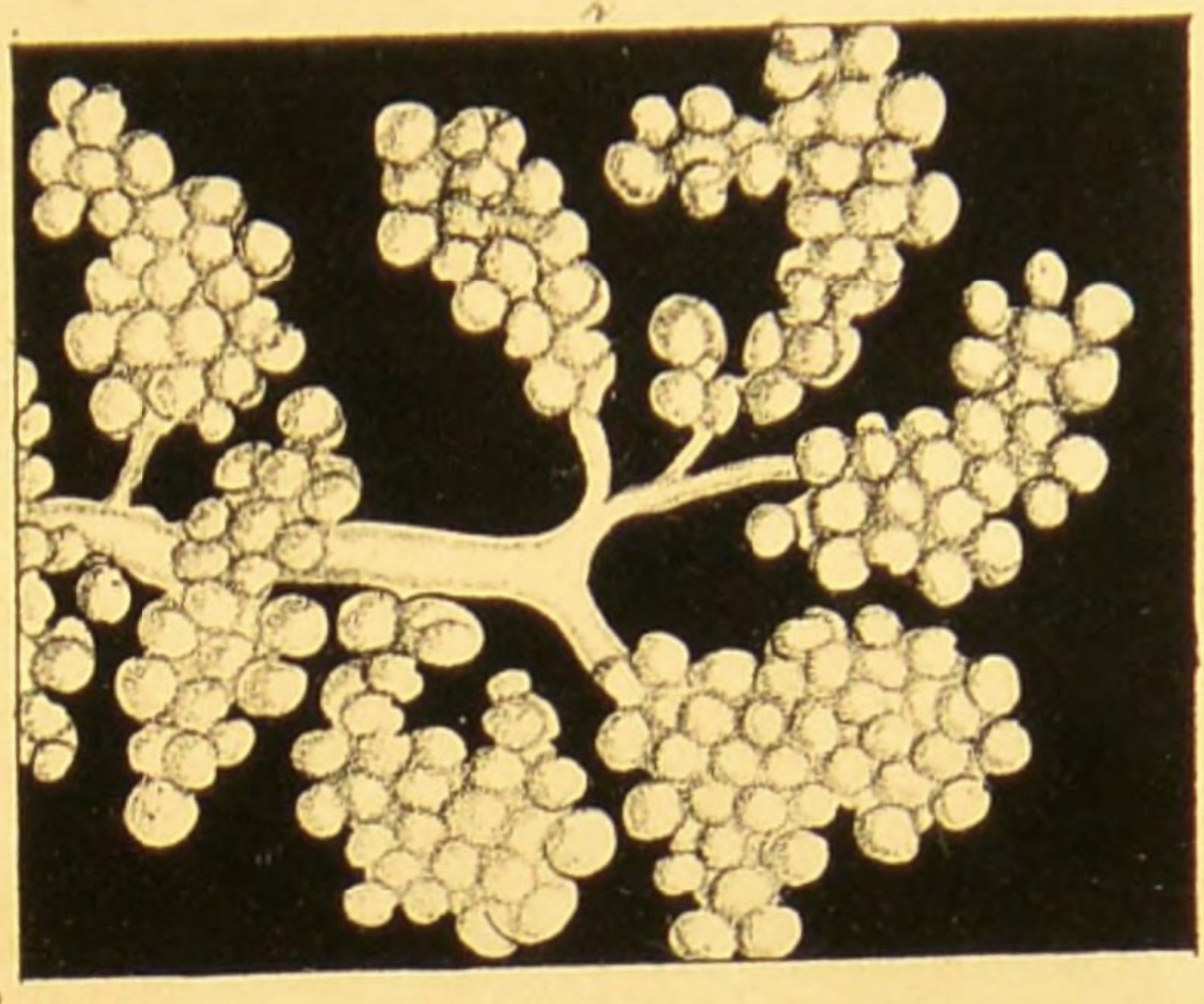 |
| Hassall's Plate LIII in Vol. 2 nicely illustrates acini and ducts of the
parotid gland. (If you follow the image-link to
the facsimile at the Wellcome Collection, note that the captions are reversed for plates LIII and LIV.)
|
A contemporary review in the Provincial Medical & Surgical Journal (1846)
reported, "The author of this work, which is appearing with commendable regularity, in monthly parts, is already favourably known to science by
his History of the British Fresh-Water Algae. The design of this present undertaking is good, and we are glad to observe the
attention of a British microscopist directed towards these objects, and to the supply of a desideratum in the medical literature of this country."
This textbook was produced fairly early in Hassall's productive career, which extended over six decades. Consult resources below for
additional biographical information.
Further resources
A B C D
E F G H
I J K L
M N O P
Q R S T
U V W X
Y Z
TOP OF PAGE / ALPHABETICAL INDEX / CHRONOLOGICAL INDEX
Clopton Havers (1657-1702)
English physician, commemorated in Haversian canals.
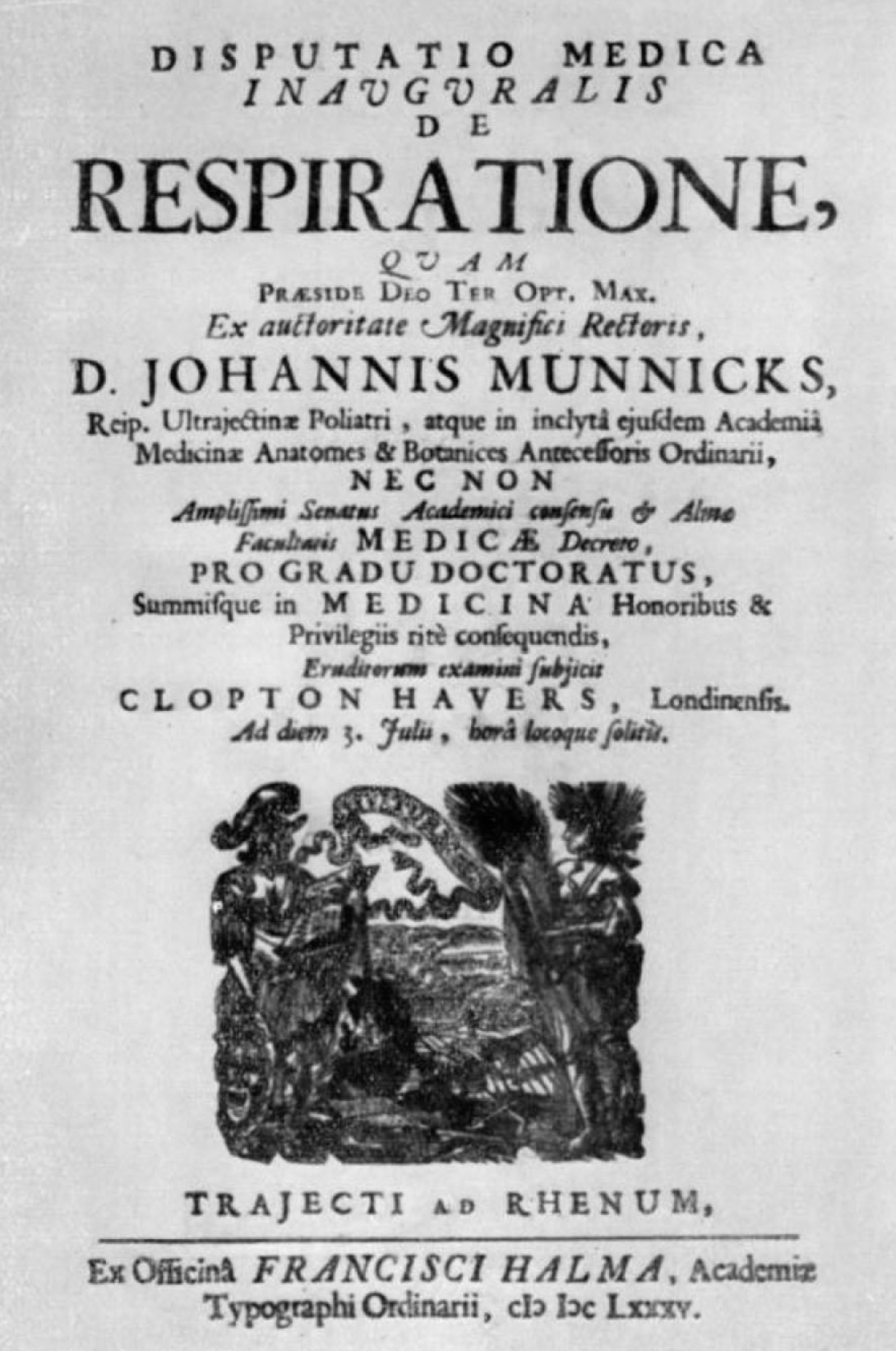
Havers studied medicine at Utrecht University; his disputation (i.e., thesis defense) "On Respiration,"
was presented there in 1685.
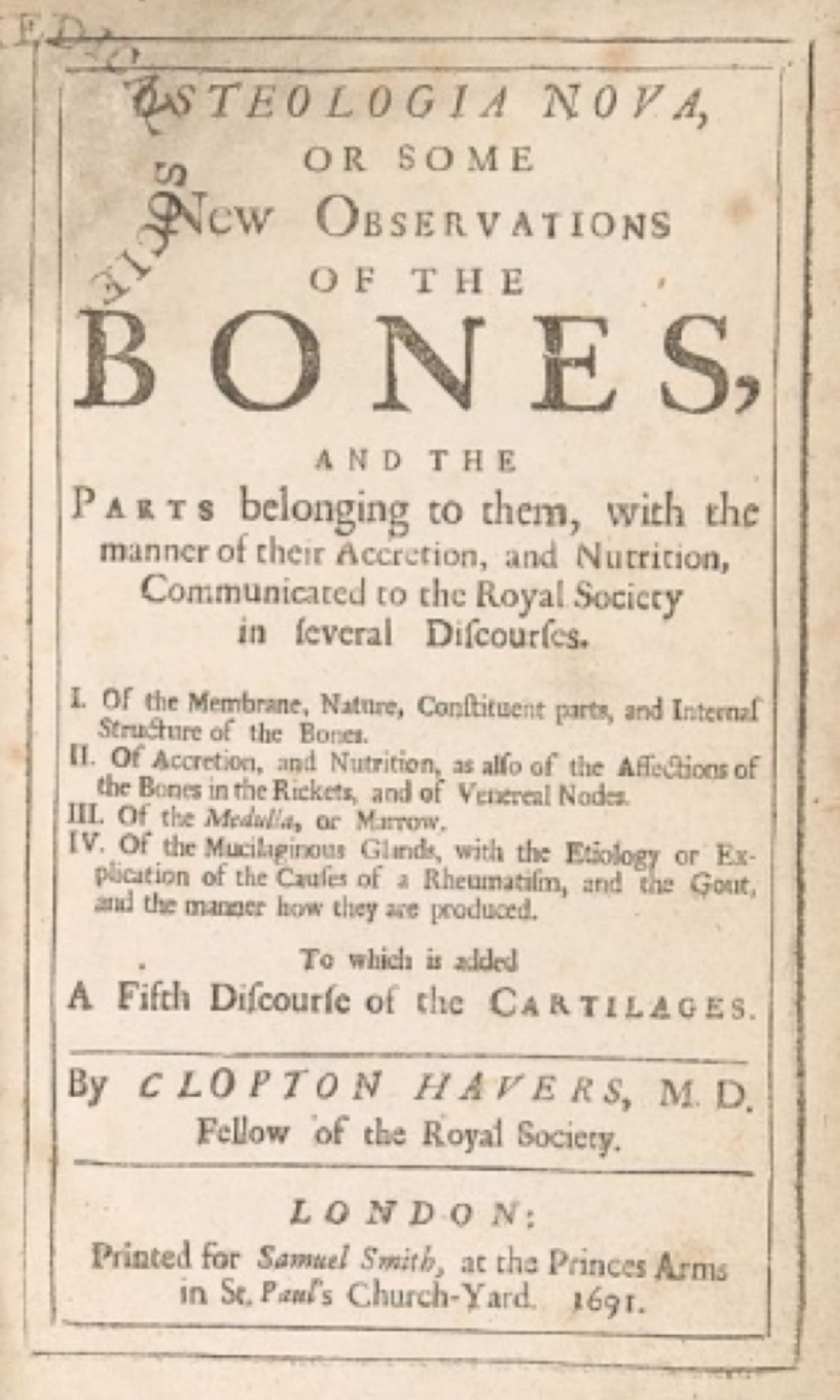
He was admitted as a Fellow of the Royal Society of London on 15 December 1686. In 1691 he presented
the work for which he is now best known, "Osteologia nova, or some new Observations of the Bones, and the Parts
belonging to them, with the manner of their Accretion and Nutrition."
Unfortunately, this volume (in Wellcome
Collection archive) lacks any illustrations of Havers' observations of microscopic anatomy of bone.
Curiously, in 1700 Dr. Clopton Havers presented to the Royal Society a report on the Chinese practice of variolation
for smallpox (i.e. vaccination). However, "There is no evidence that this initial notice excited any
attention within the medical establishment, and nothing more was heard of it in London for 13 years" [quotation
from A History of Immunology, by Arthur M. Silverstein, 2012].
Biography:
Clopton Havers, by Jessie Dobson, The Journal of Bone and Joint Surgery, vol. 34B, pp. 702-707 (1952).
Very brief bio from Wikipedia.
A B C D
E F G H
I J K L
M N O P
Q R S T
U V W X
Y Z
TOP OF PAGE / ALPHABETICAL INDEX / CHRONOLOGICAL INDEX
Hans Held (1866-1942)
German anatomist, commemorated in "calyces of Held" and "endbulbs of Held," which are exceptionally large synaptic contacts in the brainstem.
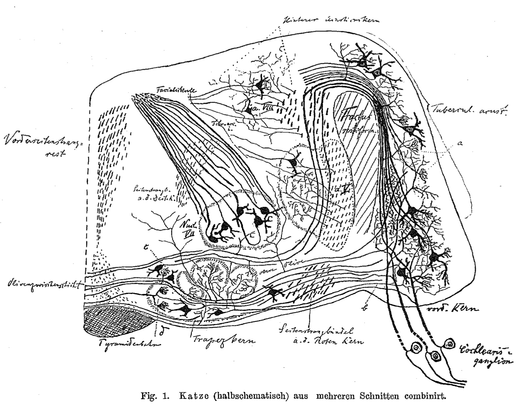 |
| The brainstem of a cat in half-section, midline at the
left side, with the calyces of Held within the trapezoid nucleus [Trapezkern] at lower left,
(beside the cross-hatched pyramidal tract). Held refers to the eponymous calyces as Faserkörben [fiber baskets]. |
The eponymous calyces and endbulbs provide rapid and reliable synaptic transmission within the auditory system. Their large size has
facilitated research into synaptic function. (For further reading, see below.)
The image at right is from: Held, H. (1893), "Die centrale Gehörleitung" [The central
auditory pathway], Arch. Anat. Physiol. Anat. 17:201-248. In this report, Held provides a thorough account of his research on auditory pathways in the
brainstem, within the limits of the histotechnology available to him:
"My investigations on the finer terminations of the cochlear fibers in the brain were based on Golgi's chromosmium silver staining as applied by
Ramón y Cajal and von Kölliker. What that first method [Weigert's Haematoxylin
myelin stain, in a previous report by Held] was unable to show, the dissolution
of axonal cylinders into terminal branches, their origin from ganglion cells, is what the silver method is able to reveal, and because this method,
under certain circumstances, distinguishes by color individual elements of a system from one another, it increases the possibility for the investigator
to clearly recognize the composition of a pathway from originally different elements. The use of this latter method, which is only capable of
extensively staining small pieces of the brain, forces the investigator, if he wants to determine and fathom a system running over long distances with
respect to the cells and fibers belonging to it, to make a composition of the details found at the individual points of the pathway in order to obtain
a uniform picture of the entire system" [p. 202, translation with some help from DeepL Translator and
Google Translate].
At the time this entry was being prepared, biographical information for Hans Held the anatomist was difficult to find on the internet;
there was no biographical entry for Held in the English-language Wikipedia. The following quote [per translation by
DeepL] is taken from a brief biography
at the German-language Wikipedia (this source also lists several of Held's publications).
"In 1888, [Held] moved to the University of Leipzig, where he received his doctorate in medicine in 1891 and became a habilitated private lecturer in 1893.
In 1899 he was appointed associate professor at the medical faculty, was 2nd prosector at the anatomical institute and became full professor of anatomy and
histology in 1917."
More on the Calyx of Held
A B C D
E F G H
I J K L
M N O P
Q R S T
U V W X
Y Z
TOP OF PAGE / ALPHABETICAL INDEX / CHRONOLOGICAL INDEX
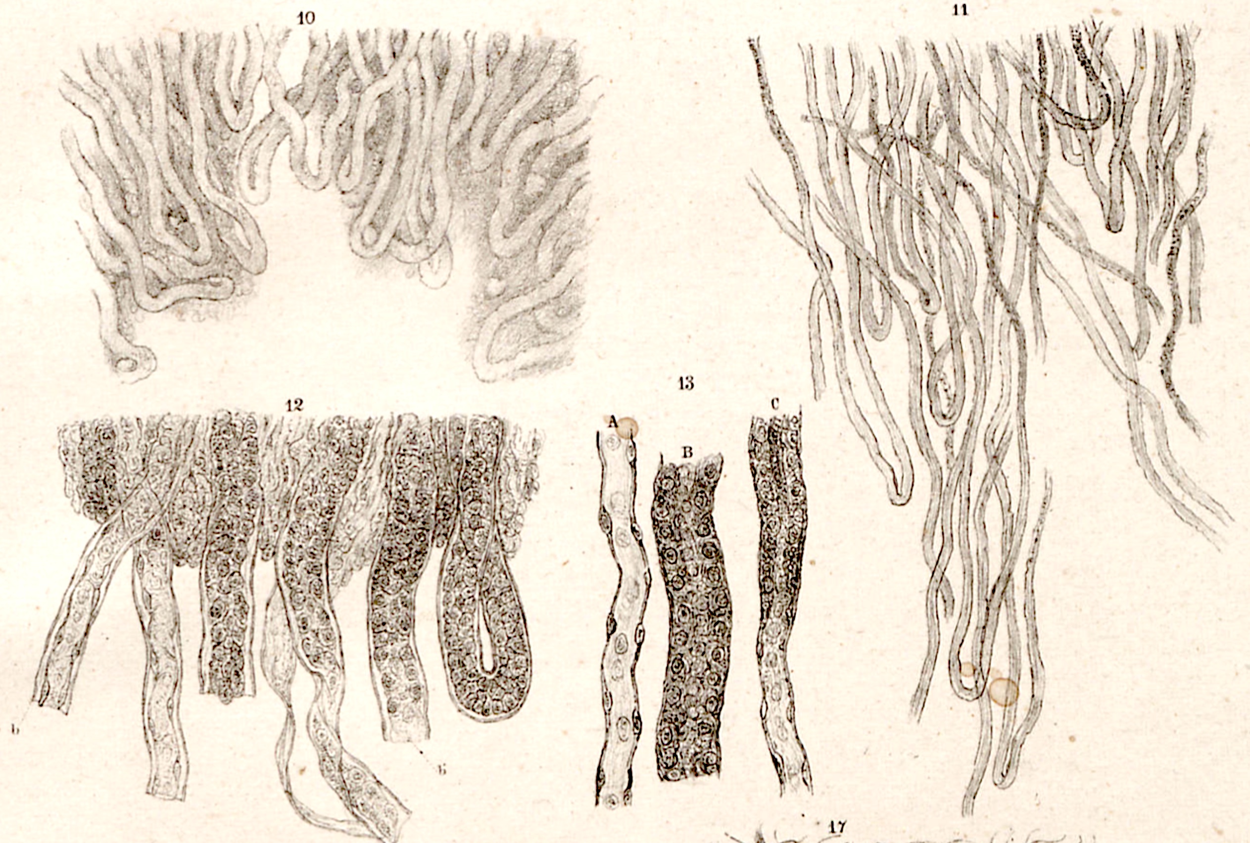 Friedrich Gustav Jakob Henle (1809-1885)
German physician, anatomist and pathologist, commemorated in the loops of Henle in the kidney and the
Henle
fiber layer in the retina, as well as numerous other anatomical structures.
After taking his doctor's degree at Bonn, Henle became a prosector for Johannes
Müller (the great comparative anatomist commemorated
in the "Müllerian duct") in Berlin. Henle worked extensively in "general anatomy," including what we would now call comparative
anatomy. During his career, he held positions in Zürich, Heidelberg, and Göttingen. While Henle was in Göttingen, his
lectures inspired at least one student, Wilhelm Waldeyer, to shift his field of study from mathematics and physics to anatomy.
(Waldeyer went on to become a renowned anatomist himself.)
In 1855 Henle released "the first instalment of his great Handbook of Systematic Human Anatomy, the last volume
of which was not published until 1873. This work was perhaps the most complete and comprehensive of its kind at that time, ...
remarkable not only for the fullness and minuteness of its anatomical descriptions but also for the number and excellence of the
illustrations" (quotation from the classic 1911 edition of Encyclopedia Britannica, available
here, at Wikisource).
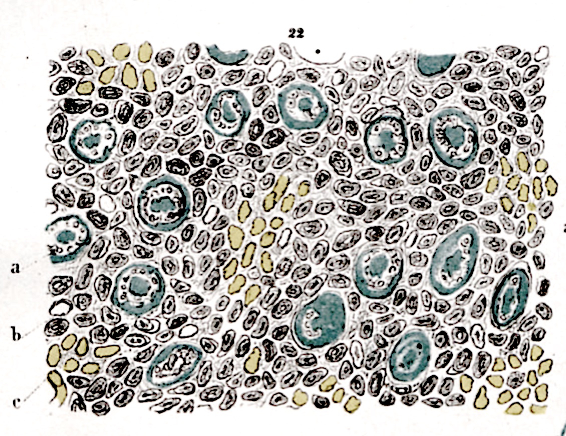 |
| Renal medullary tubules are here shown in cross-section: Collecting ducts are green, vasa recta yellow. |
Images here are from Zur Anatomie der Niere (Gottingen, 1862; accessed at
Universitätsbibliothek Heidelberg),
in which Henle described the eponymous loops of renal tubules. That the renal medulla consisted of
tubular loops had been noted a century earlier, by Exupère-Joseph Bertin, but
not until the mid-twentieth century was the counter-current function of Henle's loops
understood as essential to the concentration of urine. For an essay providing historical
context for our understanding of kidney function, from Bellini in the 1600s through Malpighi and
Bowman to the present, see "The loop of Henle as the milestone of mammalian kidney concentrating
ability: a historical review," [Koulouridis & Koulouridis, Acta Med Hist Adriat 12:413-28 (2014)], available at
PubMed or at
ResearchGate.
In additional to his anatomical studies, Henle helped to found the theory of infectious disease caused by microorganisms. His name is
associated with that of Robert Koch,
the Nobel Prize-winning bacteriologist who is remembered in "Koch's postulates."
The Wikipedia entry includes a list of numerous additional
anatomical eponyms for Henle.
An essay by Carlos Ortiz-Hidalgo, "The professor and the seamstress:
an episode in the life of Jacob Henle" (in Gaceta Médica México 2015; 151:762-9),
offers much more detail on Henle's life and relationships, both professional and personal. This essay includes a
curious note: "The story of Eliza Doolittle [in George Bernard Shaw's play Pygmalion] resembles that of Elise Egloff,
Jacob Henle's first wife." Elise's story is told in much greater detail in a German-language
Wikipedia article; this may be easily translated by copy-and-pasting
into DeepL or GoogleTranslate.
A B C D
E F G H
I J K L
M N O P
Q R S T
U V W X
Y Z
TOP OF PAGE / ALPHABETICAL INDEX / CHRONOLOGICAL INDEX
Christian Andreas Victor Hensen (1835-1924)
German physiologist, commemorated in Hensen's cells of the organ of corti in the inner
ear, also Hensen's duct (ductus reuniens) of the vestibular system and Hensen's node of the
chick embryo.
(For more on cells of Hensen, as well as on other eponyms associated with the inner ear, see
J. Schacht & J.E. Hawkins,
"A Cell by Any Other Name: Cochlear Eponyms," Audiology & Neuro Otology 2004, vol. 9, pp. 317-327, DOI: 10.1159/000081311.)
Hensen was director of the Institute of Physiology at the University of Kiel. In addition to anatomical, physiological, and
embryological studies, Hensen participated in marine biological expeditions. He is credited with coining the word "plankton."
Brief biography at Cochlear Explorers, as well
as more information on Henson's cells
Brief biography at Wikipedia.
A B C D
E F G H
I J K L
M N O P
Q R S T
U V W X
Y Z
TOP OF PAGE / ALPHABETICAL INDEX / CHRONOLOGICAL INDEX
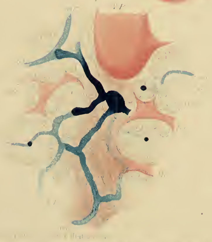 |
| Image from Hering, 1866, Ueber den Bau der Wirbelthierleber (foldout following p. 340). Bile duct injected with Prussian blue, revealing transition from bile canaliculi to an eponymous "canal of Hering." |
Karl Ewald Konstantin Hering (1834-1918)
German physiologist, commemorated in canals of Hering in the liver.
Hering's 1860 doctoral dissertation at the University of Leipzig concerned certain polychaete worms. His work on liver (image at right) was published a few years later, while he was professor at the Josephs-Akademie in Vienna:
Hering, E. (1866) Ueber den Bau der Wirbelthierleber ("On the structure of the vertebrate liver"). Archiv für mikroskopische Anatomie, 3: 88-114.
Hering went on to become "one of the greatest personalities among the German physiologists of his time" (quote from the Whonamedit entry for Hering). He studied respiratory physiology, describing the eponymous Hering-Breuer reflex in 1868. Hering is also associated with the theory of "organic memory," an idea similar to Lamarckian heredity. But his career engaged most substantially with the physiology of visual perception.
Hering is especially noted for challenging the color-vision theory of Hermann von Helmholtz. "Hering spent most of his life arguing violently with Helmholtz... Hering and Helmholtz disagreed on almost everything and the controversy lasted long after the end of both of their lives" (quote from Wikipedia entry for Hering).
Hering's son, Heinrich Ewald Hering, also became a prominent physiologist; the younger Hering is the eponym for "Hering's nerve" (the carotid branch of the glossopharyngeal nerve, responsible for the carotid sinus reflex).
A B C D
E F G H
I J K L
M N O P
Q R S T
U V W X
Y Z
TOP OF PAGE / ALPHABETICAL INDEX / CHRONOLOGICAL INDEX
Robert Hooke (1635-1703)
English polymath who made significant contributions to many areas of science. Colleague of Isaac Newton (1643-1727).
Hooke has no eponyms in histology, but he is commemorated in "Hooke's Law" of elasticity in
physics.
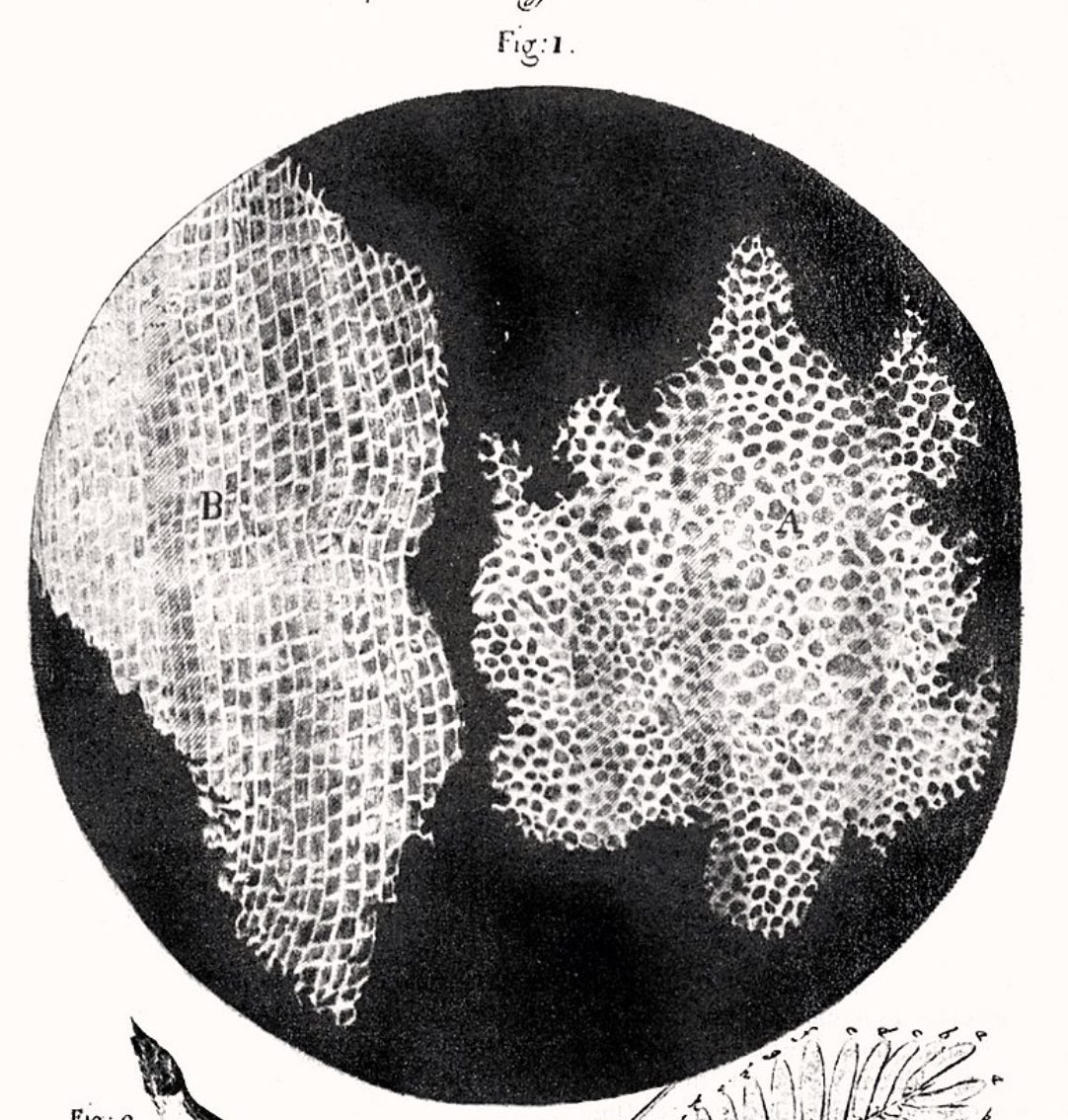
In biology, Robert Hooke's fame is due largely to
Micrographia, his 1665 account of miscellaneous observations using an
early
compound microscope. The full title is "Micrographia: or Some Physiological Descriptions of Minute Bodies Made
by Magnifying Glasses. With Observations and Inquiries Thereupon.
In Micrographia Hooke illustrated whatever was at hand, including drawings of numerous insects
and other minute animals. But it was Hooke's description of a thin slice of cork that cemented his fame as
the discoverer of cells.
The cells which Hooke drew were tiny empty chambers. But Hooke
had no understanding of cells as small bits of living material. In modern terms, Hooke's "cells" were
the cell walls that had once surrounded living cells when the cork tree was alive.
But Hooke's term "cell" was eventually transferred to the living unit during the elaboration of
Cell Theory by Schleiden and Schwann during the 1800s.
Biologists have become so accustomed to calling a unit of biological organization a "cell" that
we seldom notice that the word is an outrageous misnomer, one whose principal meaning remains that
of "small empty chamber."
A facsimile of the full, illustrated text of Micrographia may be viewed
here, at the Internet Archive.
More on Hooke from "Pioneers in Optics."
More at Wikipedia.
More at Britannica.
A B C D
E F G H
I J K L
M N O P
Q R S T
U V W X
Y Z
TOP OF PAGE / ALPHABETICAL INDEX / CHRONOLOGICAL INDEX
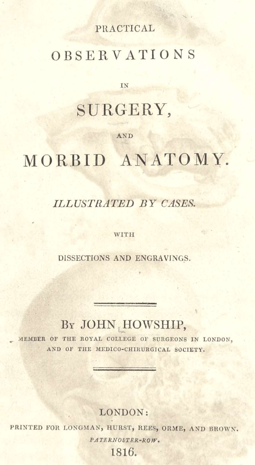 John Howship (1781-1841)
English surgeon, commemorated in Howship's lacuna, a site where
matrix is reabsorbed during bone remodelling.
Howship began his medical career as an assistant to surgeon John Heaviside, preparing demonstration
dissections of pathological anatomy specimens. Heaviside kept such specimens in a museum at his home,
for exhibition to "respectable gentlemen." Some of Howship's preparations are still preserved in the
John Heaviside's Collection at the Hunterian Museum of the Royal College of Surgeons of England.
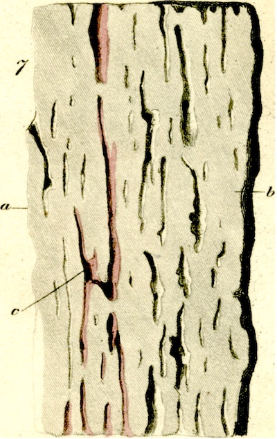 "By the time of his death, [Howship] was one of the most distinguished surgeons in England. Howship's most noteworthy
publication was his book, Practical Observations in
Surgery and Morbid Anatomy, published in 1816." The preceding quote is taken from a very brief biography
included in an article in the
Journal of Neurosurgery: Pediatrics, vol. 16, pp. 472-476 (2015), "John Howship (1781-1841) and growing skull
fracture: historical perspective," by S.C. Bir, et al.
The illustration of bone at left is from Howship's "Microscopic Observations on the Structure of Bone,"
Medico-Chirurgical Transactions,
1816, vol. 7, pp. 382-592 (doi: 10.1177/095952871600700129). This report describes in some detail
the difficulties Howship encountered during his efforts to prepare specimens of fresh bone for
microscopic examination.
The brief biography at Whonamedit.com lists additional publications by Howship.
A B C D
E F G H
I J K L
M N O P
Q R S T
U V W X
Y Z
TOP OF PAGE / ALPHABETICAL INDEX / CHRONOLOGICAL INDEX
Toshio Ito (1904-1991)
Japanese physician who discovered stellate fat-storing cells (hepatic stellate cells) in the space of Disse of the liver. Commemorated in Ito cells.
Sorting out the various cell types associated with liver sinusoids occupied much of a century; also see entry for
Kupffer.
More,
from Gastroenterology, "Ito of Ito cells," vol. 130, p. 714 (2006).
A B C D
E F G H
I J K L
M N O P
Q R S T
U V W X
Y Z
TOP OF PAGE / ALPHABETICAL INDEX / CHRONOLOGICAL INDEX
Arthur Jacob (1790-1874)
Irish ophthalmologist commemorated in "Jacob's membrane," an obsolete term for the photoreceptor layer
of the neural retina. Jacobs is considered to be the first, in 1819, to describe the retina microscopically.
The British Journal of Ophthalmology (Vol. 32, pp. 608-609, 1948) offers an eloquent biographical essay by
L.B. Somerville-Large, available
here. This essay, "The first Irish ocular-pathologist, Arthur Jacob, 1790-1874," includes
excerpts (beginning on page 608)
from Jacob's report "On the operation for the removal of cataract: as performed with a fine sewing needle through the cornea."
Jacob's description of eye surgery in an era before anesthesia, using nothing more than a pile of books and a hooked needle, is especially
poignant for those of us who have personally experienced modern lens-replacement surgery. (This was several decades before
Carl Koller introduced cocaine as a revolutionary
anesthetic for eye surgery.)
"I seat the patient in a chair and make him sit straight up or inclining, according to his height. If very tall I raise
myself by standing on a large book or two, or on anything which answers the purpose to be found at hand. In my own place of
business I find old medical folios answer the purpose well... When he is seated I lay the patient's head against my chest,
and placing the middle finger of my left hand on his lower and the forefinger on his upper eyelid, and gently holding the eye between
them, I strike the point of the needle suddenly into the cornea, about a line from its margin, and there hold it until any struggles
of the patient, which may be made, cease. There must be no hesitation here... I advise the operator to pause here for a moment,
holding the eye firmly and steadily on the point of his needle, and if necessary to say a word of encouragement or remonstrance to the patient..."
More, from Wikipedia.
A B C D
E F G H
I J K L
M N O P
Q R S T
U V W X
Y Z
TOP OF PAGE / ALPHABETICAL INDEX / CHRONOLOGICAL INDEX
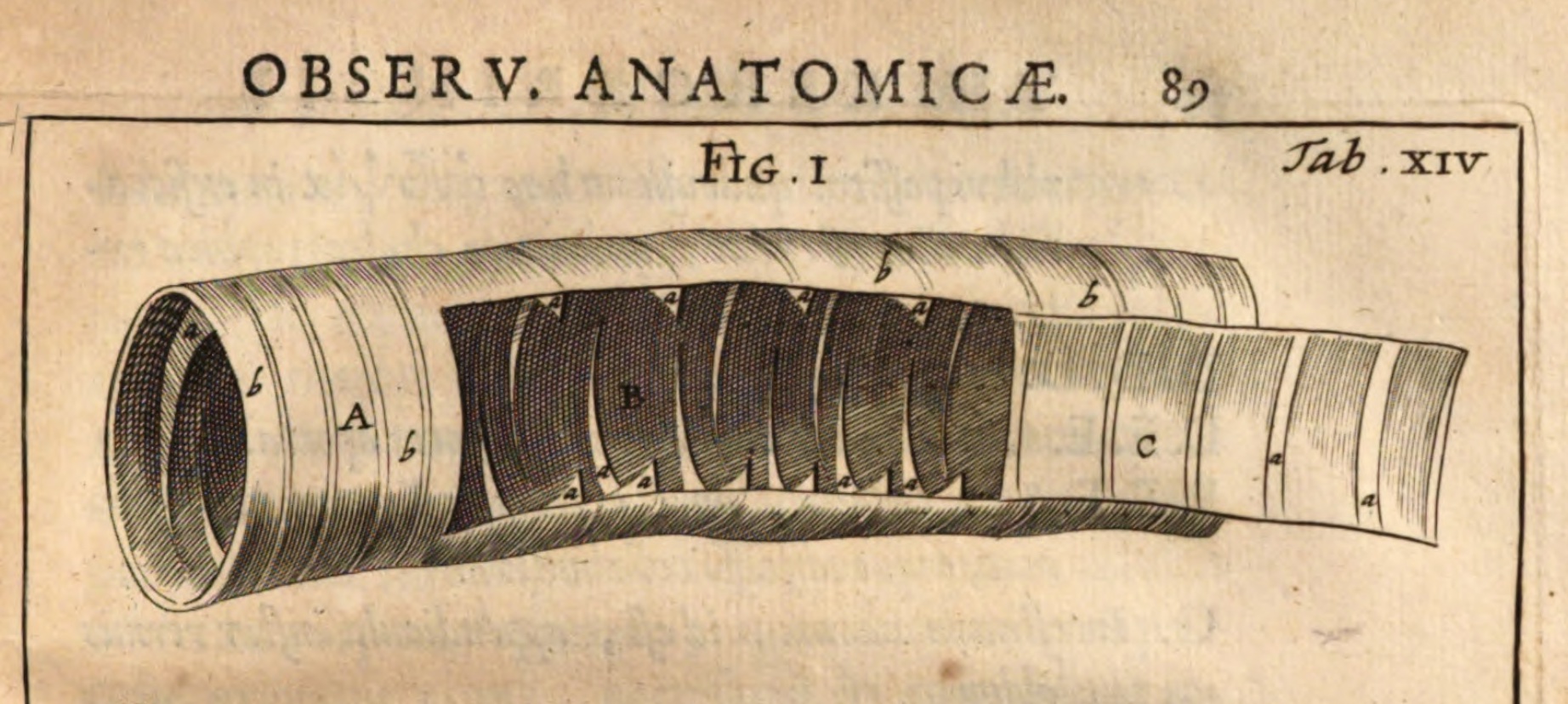 Theodor Kerckring (ca. 1638-1693)
Dutch anatomist and chemist, commemorated in valves of Kerckring (= intestinal plicae).
Kerckring studied Latin with Spinozoa in Amsterden and studied anatomy under Franciscus Sylvius (noted eponyms: Sylvian fissure and
aquaduct of Sylvius) at Leyden University. Kerckring kept a museum; he is noted for his
Spicilegium anatomicum (1670),
a collection of miscellaneous anatomical observations which includes his description of the eponymous intestinal valves.
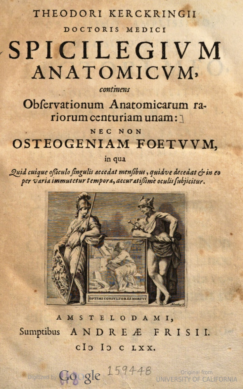
The following quotations are excerpted from "Theodore
Kerckring and His 'Spicilegium Anatomicum'," by Albert G. Nicholls,
The Canadian Medical Association Journal, pp. 480-483 (May 1940):
"The word 'Spicilegium' perhaps needs explanation. It is derived from the Latin 'Spica', a spike or head, as of flowers or grain, and in the
medical language of the day means an anthology or collection of observations which may be clinical, anatomical, or both. These observations are
presented individually on their merits and have no logical connection or sequence. Spicilegia were quite popular in Kerckring's time among
medical authors... Kerckring's Spicilegium is similar to most of the others, in that it is a sort of 'omnium gatherum' of clinical observations, rare
occurrences, anatomical notes and curiosities, and autopsy findings."
"[quoting Kerckring] 'Obs[ervation] XXXIX: In the colon and in the ileum many valves are found which, because they do not fill up the whole space,
we call valvulae conniventes.' "
"[quoting Kerckring] 'Obs[ervation] XC: A bad habit prevails in Europe of sucking the smoke of the herb Tobacco through tubes connected with it. To show in
what degree one who indulges in it very often may injure his health I submit to your attention the case of a man whom I sectioned [i.e.,
dissected] before a concourse of physicians. Being addicted to immoderate indulgence in these smoky delights he would undertake scarcely
any business without inhaling this smoke, which was, as it appeared, fatal for him... What about his trachea? Like a chimney, everywhere
covered with a black soot; the lungs dry and collapsed, almost friable.' "
Brief biography at Wikipedia.
A B C D
E F G H
I J K L
M N O P
Q R S T
U V W X
Y Z
TOP OF PAGE / ALPHABETICAL INDEX / CHRONOLOGICAL INDEX
Wilhelm Kiesselbach (1839-1902)
German otolaryngologist, commemorated in Kiesselbach's area, a frequent site for nosebleeds.
After studying medicine in Göttingen, Marburg and Tübingen, Kiesselbach moved to the University of Erlangen where he
remained for the remainder of his career, eventually becoming director of otolaryngology at the university clinic. Although Kiesselbach
specialized in otology, he also actively pursued research in rhinology and laryngology. According to Deutsche Biographie, Kiesselbach was widely acclaimed for his 1891 publication, Die galvanische Reaktion
der Sinnesnerven [The galvanic reactions of the sensory nerves].
Kiesselbach described the eponymous feature of the nasal septum in 1884, in Über spontanes Nasenbluten [About spontaneous nosebleeds],
Berliner Klinische Wochenschrift 22, 1884.
(Kiesselbach's area is also known as "Little's area." Severe nasal bleeding originating from this area had been previously
described by James Little in 1879, in The Hospital
Gazette, v. 6(1): 5-6.)
German-language entry for Kiesselbach at Deutsche Biographie.
Brief entry for Kiesselbach at Wikipedia, with a short bibliography.
Very brief note for Kiesselbach at the blog, Medical Terminology Daily.
A B C D
E F G H
I J K L
M N O P
Q R S T
U V W X
Y Z
TOP OF PAGE / ALPHABETICAL INDEX / CHRONOLOGICAL INDEX
David G. King (b. 1948)
Obscure American neurobiologist and evolutionary geneticist, with no eponyms. King has created a popular
hypertext histology resource which self-indulgently includes his own entry right
here on this "Eponyms and historical notes" page.
Yes, this is ME -- the author of this website.
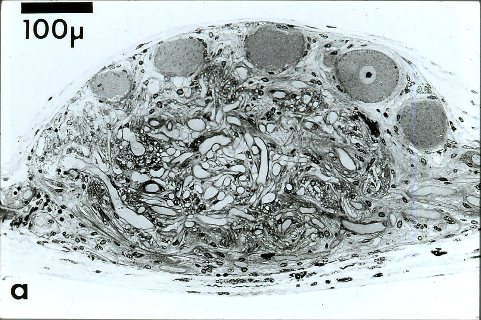
King's somewhat-esoteric histological works include descriptions of synaptic organization
in a lobster ganglion; of peculiar nerve fibers in flies; and of a specialized region
of the digestive tract in flies (respectively, 1976a,b;
1980,
1983a,b; and 1988,
1989, 1991).
King has also characterized genetic sites of repetitive DNA as
"evolutionary tuning knobs"
and has speculated on the evolutionary basis for
individually-identifiable nerve cells. In one recent publication (2024)
King addressed a formerly-heretical perspective on mutation.
These diverse topics -- none of which is part of the working vocabulary for most biologists -- have all been inspired
by his appreciation for (as Marcello Malpighi wrote over three hundred years ago) "extremely minute parts
so shaped and situated as to form a marvelous organ." His efforts have been further united by his desire to understand the
patterns of structure and genetics that enable the evolutionary malleability of animal behavior.
Curiously, King's academic ancestry includes Rudolf Virchow, along a couple different branches.
(Academic genealogies, mapping student/mentor relationships, can be viewed at The
Academic Family Tree. King's own tree can be found by choosing "neuroscience"
and entering his name, "David G. King," in the search box.)
King's personal webpage at Southern Illinois University Carbondale
includes an annotated list of publications as well as some brief
autobiographical notes and links to his
nature photography.
A B C D
E F G H
I J K L
M N O P
Q R S T
U V W X
Y Z
TOP OF PAGE / ALPHABETICAL INDEX / CHRONOLOGICAL INDEX
August Köhler (1866-1948)
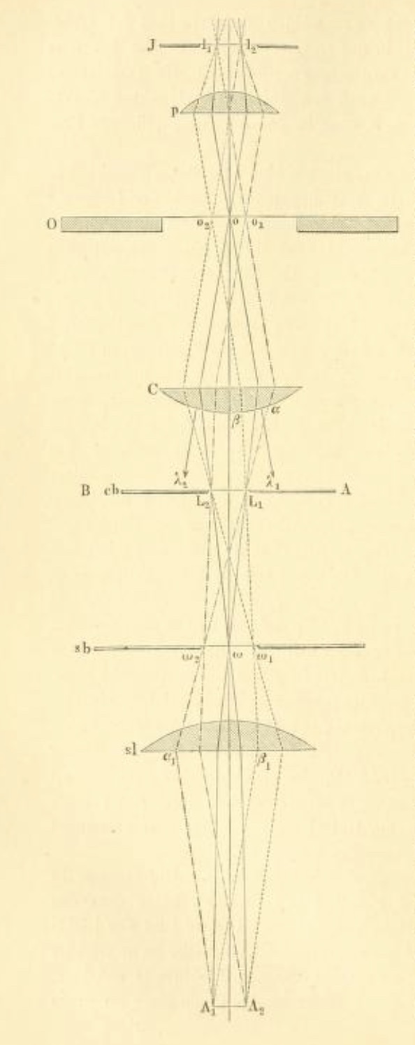
German biologist and physicist, inventor of "Köhler illumination," an optical method
for uniformly illuminating a microscopic specimen.
Köhler's doctoral research at the University of Giessen, studying the taxonomy of
limpets (a type of gastropod mollusc),
depended on high quality photomicrography. This effort was hampered by lack of a satisfactory
means for providing bright, uniform illumination to the microscope's field of view.
To overcome this problem, Köhler developed an optical configuration, now known as
"Köhler illumination," which he published in
Zeitschrift für wissenschaftlichen Mikroskopie (v. 10: pp. 433-440) in 1893,
the same year that he received his doctorate.
"Yet, other researchers in the field did not immediately realize the significance and importance
of the Köhler illumination methodology. In fact, ... Köhler's name seemed as if it
might vanish into obscurity as he left the University of Geissen to work as a grammar school teacher
in Bingen, Germany."
[quote from a much more extensive biography of Köhler at
Pioneers in Optics, from Florida State University.]
In 1900, however, Köhler was hired by the Zeiss Optical Works where he continued work improving the tools and
techniques of microscopy. His invention has since been widely adopted by microscopists everywhere.
Explanation of optics for Köhler illumination,
from "Optical Microscopy Primer."
This website is an excellent resource for understanding microscopes and their history.
Also see "Pioneers in optics" for more
on the history of optics and microscopy
Simplified explanation of Köhler optics
from Wikipedia.
Köhler's
original publication, in German.
English translation of Köhler's original publication.
[More about Köhler, from Wikipedia]
A B C D
E F G H
I J K L
M N O P
Q R S T
U V W X
Y Z
TOP OF PAGE / ALPHABETICAL INDEX / CHRONOLOGICAL INDEX
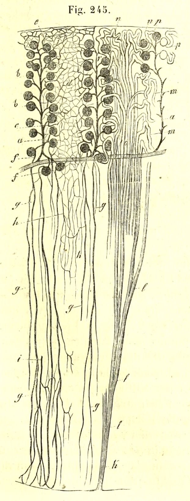 |
| Essential microanatomy of kidney, from Kölliker's Handbuch der Gewebelehre. Vasculature is shown on left side,
epithelial tubules on right. (Loops of Henle were not yet understood in 1852.) |
Rudolph Albert von Kölliker (1817-1905)
Swiss zoologist / embryologist / anatomist / physiologist. Kölliker has been called the "father of modern histology" (e.g.,
[1]). Note "modern" in this appellation; Kölliker should not be confused with Bichat, an earlier
"father of histology" who had previously defined a foundation for tissue studies but who did not himself use a microscope. In spite of Kölliker's
stature, eponyms commemorating his discoveries are rather obscure:
Kölliker's organ in the developing inner ear [2] and
Kölliker's organs in baby octopus.
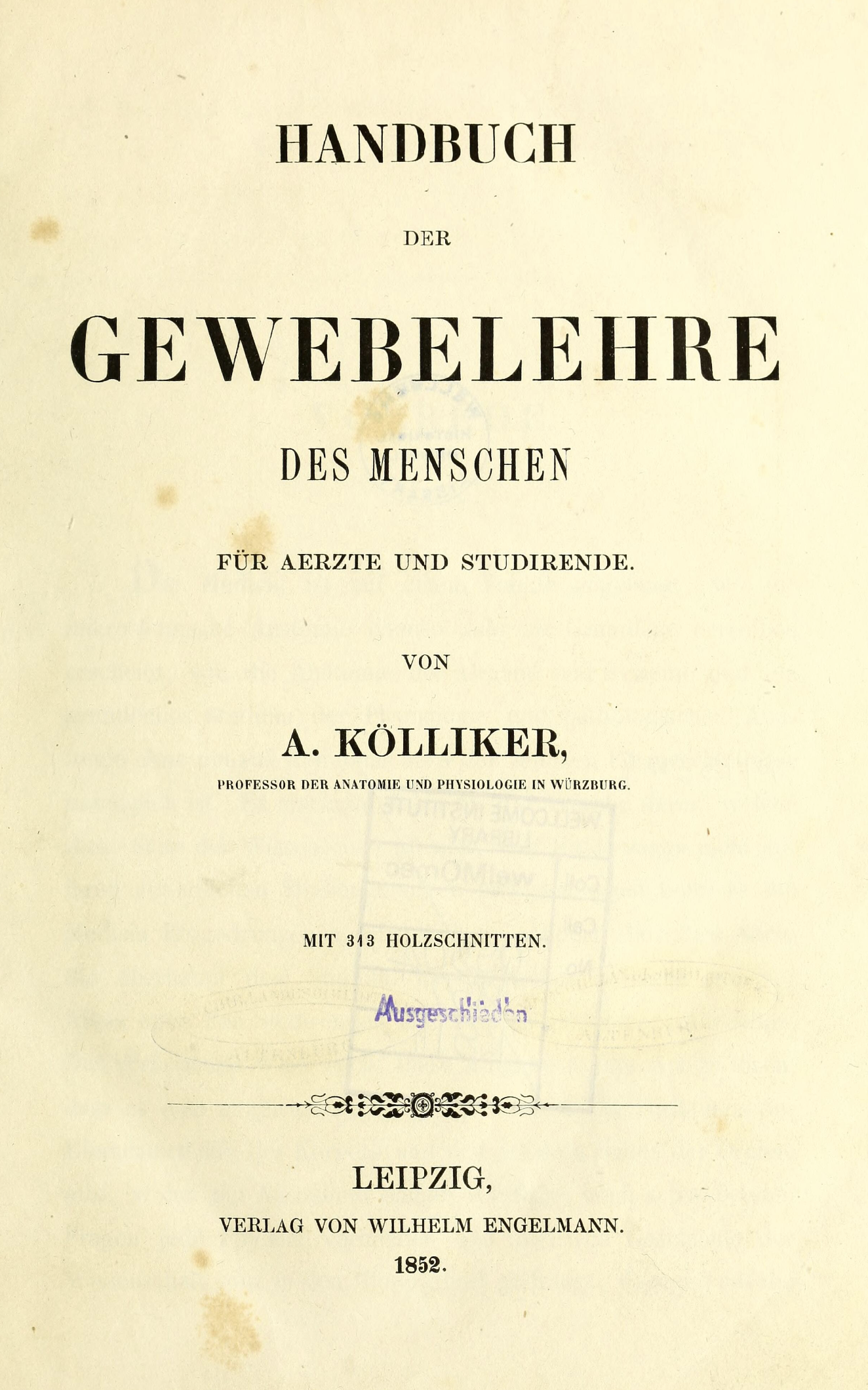
In 1852, Kölliker published a comprehensive textbook, Handbuch der
Gewebelehre des Menschen (translated into English in 1854 as Manual of human histology). This text continued
through several editions (the 6th in 1902) and served for generations to define the field of study now known as histology. The 6th edition
is also a rare historical resource; a review in Nature of the 6th edition
declared, "Too much praise cannot be given to the bibliographical notices, which are far more complete than are to be found in any other work on
histology."
Note that Kölliker used the vernacular "Gewebelehre" (literally, "tissue-teaching") rather than the German-language alternative
"Histologie" that had been introduced in 1819 by Mayer's text Ueber Histologie.
Unlike the authors of prior texts such as Mayer (1819) and Hassall (1849),
Kölliker emphasized cells as the basis for tissue structure.
Kölliker's place in the history of histology is nicely captured in the following excerpts from the classic 1911 edition of The Encyclopedia Britannica
[3]:
"Kölliker's name will ever be associated with that of the tool with which during his long life he so assiduously and successfully worked,
the microscope. The time at which he began his studies coincided with that of the revival of the microscopic investigation of living
beings. Two centuries earlier the great Italian Malpighi had started, and with his own hand had carried far the
study by the help of the microscope of the minute structure of animals and plants. After Malpighi this branch of knowledge, though
continually progressing, made no remarkable bounds forward until the second quarter of the 19th century, when the improvement of the
compound microscope on the one hand, and the promulgation by Theodor Schwann and Matthias Schleiden of the
"cell theory" on the other, inaugurated a new era of microscopic investigation.
"Into this new learning Kölliker threw himself with all the zeal of youth, wisely initiated into it by his great teacher
Henle... But Kölliker had another teacher besides Henle, the even greater Johannes Müller,
whose active mind was sweeping over the whole animal kingdom, striving to pierce the secrets of the structure of living creatures
of all sorts, and keeping steadily in view the wide biological problems of function and of origin, which the facts of structure might
serve to solve. We may probably trace to the influence of these two great teachers, strengthened by the spirit of the times,
the threefold character of Kölliker's long-continued and varied labours...
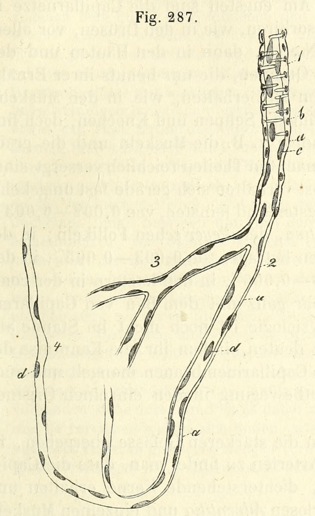 |
| Caption from translation of Handbuch der Gewebelehre: "Finest vessels on the arterial side of the
capillaries. 1, an artery of the smallest size; 2, transitional vesssel; 3, coarser capillary; 4, finer capillary: a,
structureless coat, with a few nuclei, representing the t. adventitia; b, nuclei of the muscular fibre-cells; c,
nuclei within the minute artery, probably still belonging to an epithelium; d, nuclei of the capillaries and transitional
vessels."
|
"He was among the first, if not the very first, to introduce into [embryology] the newer microscopic
technique -- the methods of hardening, section-cutting and staining. By doing so, not only was he enabled to make rapid
progress himself, but he also placed
in the hands of others the means of a like advance.
"But neither zoology nor embryology furnished Kölliker's chief claim to fame. If he did much for these branches of science,
he did still more for histology, the knowledge of the minute structure of the animal tissues. This he made emphatically his own.
It may indeed be said that there is no fragment of the body of man and of the higher animals on which he did not leave his mark, and in
more places than one his mark was a mark of fundamental importance. Among his earlier results may be mentioned the demonstration
in 1847 that smooth or unstriated muscle is made up of distinct units, of nucleated muscle-cells... A few years before this men were
doubting whether arteries were muscular, and no solid histological basis as yet existed for those views as to the action of the nervous
system on the circulation, which were soon to be put forward, and which had such a great influence on the progress of physiology.
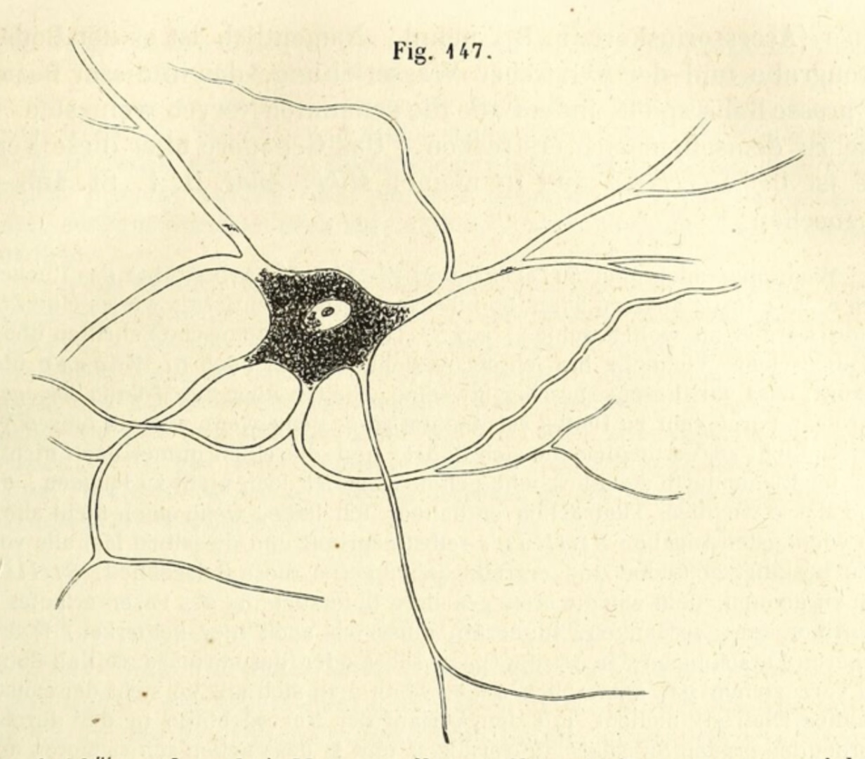 |
| Nerve cell, from Handbuch der Gewebelehre. |
"Even to enumerate, certainly to dwell on, all his contributions to histology would be impossible here... The results at which he
arrived were recorded partly in separate memoirs, partly in his great textbook on microscopical anatomy, which first saw the light in 1850,
and by which he advanced histology no less than by his own researches. In the case of almost every tissue our present knowledge contains
something great or small which we owe to Kölliker; but it is on the nervous system that his name is written in largest letters.
So early as 1845, while still at Zürich, he supplied what was as yet still lacking, the clear proof that nerve-fibres are continuous
with nerve-cells, and so furnished the absolutely necessary basis for all sound speculations as to the actions of the central nervous system...
"Naturally a man of so much accomplishment was not left without honours. Formerly known simply as Kölliker, the title "von"
was added to his name. He was made a member of the learned societies of many countries; in England, which he visited more than once,
and where he became well known, the Royal Society made him a fellow in 1860, and in 1897 gave him its highest token of esteem, the Copley
medal."
To offset somewhat the patently hagiographic tone of the above account from Encyclopedia Britannica, it might be mentioned here that
Kölliker was critical of Darwinism and supported a non-Darwinian view of evolutionary processes [4].
Works by Kölliker
Kölliker, A. Handbuch der Gewebelehre des Menschen, 1852.
Kölliker, A. Manual of human microscopical anatomy,
1854, translation by George Busk and Thomas Henry Huxley
Kölliker, A. Entwicklungsgeschichte des Menschen und der höheren
Thiere. Leipzig: Engelmann; 1879.
Partial collection of additional works by A. Kölliker, at
Biodiversity Heritage Library.
Additional information
Albert von Kölliker (1817-1905) Würtzburger Histologist.
pdf at JAMA. 1968;206(9):2111-2112. doi:10.1001/jama.1968.03150090187031
Brief biography at Wikipedia.
Citations noted above
[1] "The history of radial glia," by Marina Bentivoglio & Paolo Mazzarello, Brain Research Bulletin, Vol. 49, p. 305, 1999 (https://doi.org/10.1016/S0361-9230(99)00065-9).
[2] "Kölliker's Organ and the Development of Spontaneous Activity in the Auditory System," by M. W. Nishani Dayaratne et al, BioMed Research International
http://dx.doi.org/10.1155/2014/367939
[3] The classic 11th edition of The Encyclopedia Britannica (1911) is in public domain and accessible through several online sources, such as
Project Gutenberg and
Wikisource.
[4] Citations for Kölliker's non-Darwinian view are provided in the Kölliker entry in
Wikipedia.
A B C D
E F G H
I J K L
M N O P
Q R S T
U V W X
Y Z
TOP OF PAGE / ALPHABETICAL INDEX / CHRONOLOGICAL INDEX
 Wilhelm Krause (1833-1910)
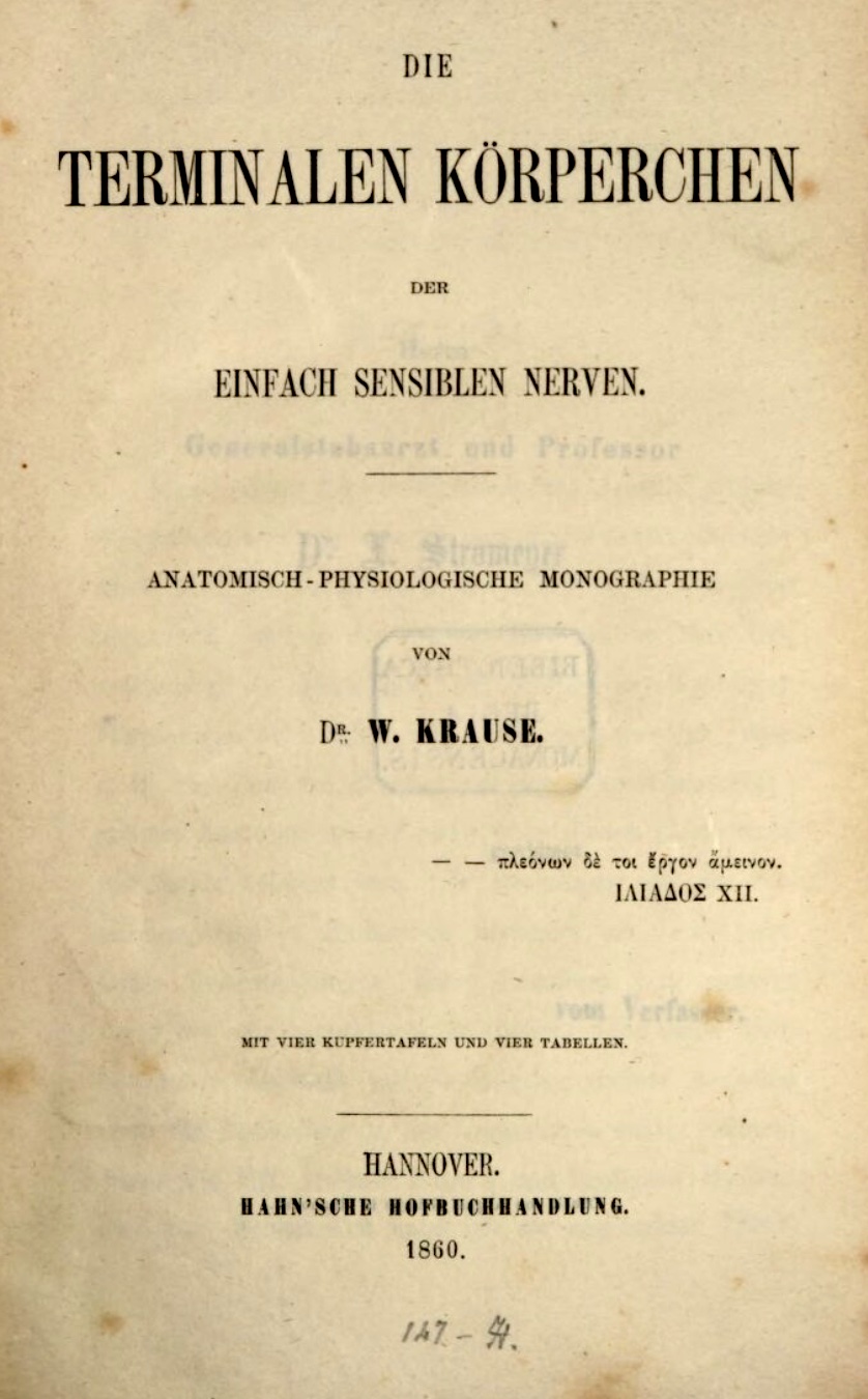
German anatomist, commemorated in endbulbs of Krause (mucocutaneous receptors).
Unlike receptor cells in special sense organs, "the receptors in or beneath the surface
of the skin were generally named after those who first described them (e.g., Golgi tendon organs, Krause
end-bulbs, Meissner's corpuscles, Merkel discs, Pacinian
corpuscles, and Ruffini cylinders)" ["Receptor
Visionaries," by Nicholas Wade, Perception, 47: 833-850 (2018)].
Selected publications by Krause:
Brief biography at Wikipedia.
A B C D
E F G H
I J K L
M N O P
Q R S T
U V W X
Y Z
TOP OF PAGE / ALPHABETICAL INDEX / CHRONOLOGICAL INDEX
Karl Wilhelm von Kupffer (1829-1902)
German anatomist whose major work was in embryology and neuroanatomy, but who is commemorated in Kupffer cells of the liver.
While using a gold chloride stain to find nerve fibers in the liver, Kupffer observed stellate cells, which he described
in 1876 as "Sternzellen" (literally, "star-cells"), associated with liver sinusoids. In 1898, he reported that
these cells could take up carbon particles from India ink injected into blood vessels. Sorting out the identities of several
sinusoid-associated cell types -- including liver macrophages (now known as Kupffer cells), the vitamin-storing stellate cells
(now known as Ito cells), and hepatic sinusoidal endothelial cells -- took several decades.
Some of this history is reported in some detail, along with biographical information, in "Karl
Wilhelm Kupffer And His Contributions To Modern Hepatology," Comparative Hepatology (2004), by Kenjiro Wake.
Selected publications by Kupffer
- "Ueber Sternzellen der Leber. Briefliche Mitteilung an Prof. Waldyer" [About stellate cells of the liver. Letter to Prof. Waldyer].
Arch mikr Anat 1876, 12:353-358.
- "Ueber die sogennanten Sternzellen der
Säugethierleber" [About the so-called stellate cells of the mammalian liver]. Arch mikr Anat 1899, 54:254-288.
Brief biography at Wikipedia.
A B C D
E F G H
I J K L
M N O P
Q R S T
U V W X
Y Z
TOP OF PAGE / ALPHABETICAL INDEX / CHRONOLOGICAL INDEX
Karl Langer (1819-1887)
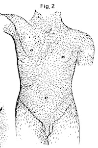
Austrian anatomist, commemorated in Langer's lines.
Click on image for a more complete diagram of the "cleavability of the cutis," as illustrated by Langer.
Langer's 1861 description of Langer's lines is fascinating;
it may found here:
"On the anatomy and physiology of the skin. I. The cleavability of the cutis," British Journal of Plastic Surgery, v. 31 pp. 3-8, 1978.
[Translated from Langer, K. (1861). "Zur Anatomie und Physiologie der Haut. I. Uber die Spaltbarkeit der Cutis,"
Sitzungsbericht der Mathematisch-naturwissenschaftlichen Classe der Kaiserlichen Academic der Wissenschaften, v. 44, p. 19.]
More on Langer's lines, from Wikipedia.
Brief bio from Wikipedia.
A B C D
E F G H
I J K L
M N O P
Q R S T
U V W X
Y Z
TOP OF PAGE / ALPHABETICAL INDEX / CHRONOLOGICAL INDEX
Paul Langerhans (1847-1888)
German histologist, commemorated in islets of Langerhans (= pancreatic islets) and Langerhans
cells of epidermis.
Eponym confusion can have serious consequences! SEE
Langhans BELOW. Langerhans cells should
not be conflated with Langhans cells (in granulomas).
Langerhans attended school in Jena, where he was a pupil of Ernst Haeckel.
He subsequently worked at the Berlin Pathological Institute in the laboratory of Rudolf Virchow (the "father of
pathology") who became a close friend. In 1868, while still a medical student,
Langerhans described the dendritic epidermal cells which now bear his name and which he
regarded as sensory. (It took over a century before the immunological function of Langerhans cells was appreciated.)
Shortly after, in his 1869 dissertation, Langerhans described cells forming "roundish little heaps" (rundlichen Häuflein)
in the pancreas of rabbits and other animals; these cell clusters were named "ilots de Langerhans" in 1893 by the French histologist
G.-E. Languesse, who described them in humans.
It remained for other researchers to work out the endocrine nature of these cell clusters. (Oskar Minkowski and Joseph von
Mering famously induced diabetes in 1889, by surgically removing a dog's pancreas. This experiment had already been performed
more than two centuries earlier by Johann von Brunner, but Brunner failed to connect the symptoms which his
operation produced with the disease of diabetes.)
Langerhans also contributed to studies on pathology of tuberculosis; he was forced to retire to the
island of Madeira after contracting the disease himself. During his years in Madeira he continued to practice medicine as well as pursue studies
on comparative and invertebrate anatomy, until his early death at age 40.
"Contributions to the microscopic anatomy of
the pancreas" (a reprint, with complete translation by H. Morrison, of Langerhans' Beiträge zur mikroskopischen Anatomie der Bauchspeicheldrüse, 1869)
includes a splendid introductory essay by Morrison describing Langerhans' pancreatic research as well as offering considerable biographical detail.
(This translation and original essay were published in the Bulletin of the Institute of the History of Medicine,
Vol. 5, pp. 259-297, 1937.)
Brief biography from Journal of Clinical Pathology,
vol. 55, p. 243 (2002), by S. Jolles. For more, see the
citation above.
Brief biography from Diabetes, vol. 1, pp. 411-413
(1952), by J.H. Barach.
Additional publications by Langerhans, from Deutsche Biographie.
Brief biography from Wikipedia.
A B C D
E F G H
I J K L
M N O P
Q R S T
U V W X
Y Z
TOP OF PAGE / ALPHABETICAL INDEX / CHRONOLOGICAL INDEX
Theodor Langhans (1839-1915)
German pathologist, commemorated by Langhans giant cells
in granulomas and the layer of Langhans (the cytotrophoblast of the placenta).
Eponym confusion can have serious consequences! The name Langhans should not be confused with
the more familiar name of Langerhans (of pancreatic islet fame). Regarding Langhans cells and Langerhans cells, "The terms are not infrequently mixed up in journal articles, textbooks, and histology reports" [ 1 ]. In at least two cases, confusion between Langerhans and Langhans has led to inappropriate treatment for a misdiagnosed disease [ 2 ].
[1] Zhang and Cadogan (Nov. 3, 2020) Life in the Fast Lane.
[2] Pritchard et al (2003) The Lancet, 362: 299.
Langhans studied medicine in Heidelberg, Göttingen, Berlin, and Würtzburg; his mentors included Henle and Virchow. The eponymous giant cells are described in: Langhans, T. (1868) Ueber Riesenzellen mit wandständigen Kernen in Tuberkeln und die fibröse Form des Tuberkels [On giant cells with peripheral(?) nuclei in tubercles and the fibrous form of the tubercle]. Archiv für pathologische Anatomie und Physiologie und für klinische Medicin 42: 382-404.
Additional publications are listed in Zhang and Cadogan (Nov. 3, 2020) Life in the Fast Lane.
A brief biography / eulogy is available in German, which may be easily translated by copy-and-pasting into
GoogleTranslate.
A B C D
E F G H
I J K L
M N O P
Q R S T
U V W X
Y Z
TOP OF PAGE / ALPHABETICAL INDEX / CHRONOLOGICAL INDEX
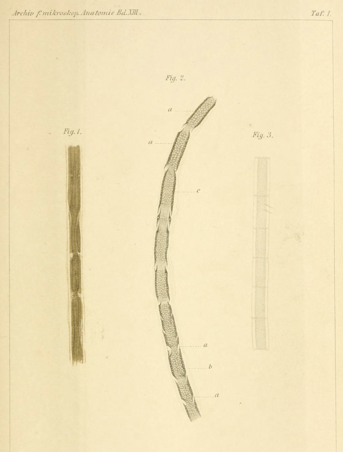 |
Image accessed at
the Internet Archive |
A. J. (Ai John) Lanterman (1845-1898)
American surgeon, commemorated by Schmidt-Lanterman clefts in myelin.
Lanterman graduated from Columbia University College of Physicians and Surgeons in 1868, then travelled to Europe for further professional development. In 1874, while completing his studies with Wilhelm Waldeyer in Strasbourg, Germany, Lanterman reported a preliminary notice on the eponymous feature of myelin. (In the same year, this same structure was reported independently by Schmidt.) Lanterman's description was subsequently published in 1877 as Über den feineren Bau der markhaltigen Nervenfasern ("On the finer structure of myelinated nerve fibers") in Archiv für mikroskopische Anatomie, 13:1-8 (image at right). Apart from this contribution during his time with Waldeyer, Lanterman evidently had little if any impact on the field of histology. (Waldeyer himself was an anatomist of some importance, one who gave us the words "neuron" and "chromosome.")
In 1878, after returning to America and serving briefly as professor of anatomy at Bellevue Hospital in New York, Lanterman moved to Colorado where he practiced medicine and acquired "very valuable mining interests in the Cripple Creek district" (quote from a Colorado newspaper, The Salida Mail, Dec. 13, 1898). Lanterman is buried in Youngstown, Ohio, where he was born and where his paternal grandparents owned several coal mines.
Detailed information on Lanterman's professional career seems to be quite scarce on the internet. My principal starting point for this entry was the Polish Wikipedia with its included links.
A B C D
E F G H
I J K L
M N O P
Q R S T
U V W X
Y Z
TOP OF PAGE / ALPHABETICAL INDEX / CHRONOLOGICAL INDEX
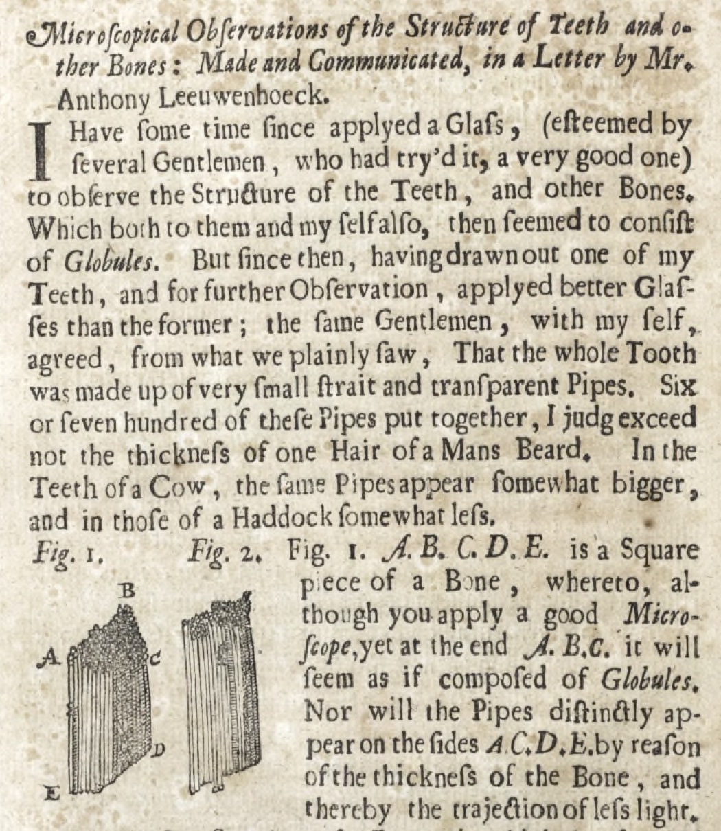
Antonie van Leeuwenhoek (1632-1723)
Dutch businessman (he ran a drapers shop) and amateur scientist, elected into the Royal Society of London in 1680.
Although not commemorated in any familiar histological eponyms, Leeuwenhoek is known as the "Father of Microbiology" for his discoveries of living things too small
to be seen with the unaided eye.
Leeuwenhoek used powerful single-lens instruments which
he made himself. Compound microscopes (based on the principle
of two lenses: an objective lens which projects a magnified image that is then magnified further by an eyepiece lens) had already been invented
and applied to good effect by researchers such as Robert Hooke. Compound microscopes were potentially more
powerful as well as easier to use than Leeuwenhoek's simple lenses, but Leeuwenhoek proved uniquely able to apply his simple instruments
to make momentous discoveries. He described these discoveries in many letters to the Royal Society of London, where his letters
were translated into English or Latin by Henry Oldenburg
(1618-1677, first secretary to the Royal Society), who introduced the famous term "animalcules" in these translations. Included
among these letters were Leeuwenhoek's brief descriptions of teeth and bone:
"Microscopical observations of the structure of teeth and other
bones" (1675), Phil. Trans. vol. 12, pp. 1002-1003.
More on Leeuwenhoek from "Pioneers in Optics."
Biography from Wikipedia.
A B C D
E F G H
I J K L
M N O P
Q R S T
U V W X
Y Z
TOP OF PAGE / ALPHABETICAL INDEX / CHRONOLOGICAL INDEX
Franz von Leydig (1821-1908)
German zoologist and comparative anatomist, commemorated in Leydig cells (interstitial
cells of testis).
Leydig also first described lipocartilage in 1854, only recently (re)discovered by Raul Ramos et al. (For a nice summary of this rediscovery, see Science 387:136-137, 9 Jan. 2025)
The remainder of this entry is largely gleaned from "Franz von Leydig (1821-1908), pioneer of
comparative histology" [M. Schneider, 2012, Journal of Medical Biography, vol. 20, pp, 79-83; doi: 10.1258/jmb.2011.011013)].
The interested reader is encouraged to access this paper at an academic library.
Leydig received his doctorate in Würzburg, where he subsequently taught microscopic anatomy under the supervision
of Albert von Kölliker. He later served in
positions in Tübingen and Bonn.
Leydig's interest in natural history began in childhood. His first microscope, at age 12, had lenses held by wooden and cardboard fittings.
His lifelong career in zoological research, including numerous studies of a wide range of invertebrate as well as vertebrate animals, is surveyed in his last book,
Horae Zoologicae," published in 1902.
The book generally regarded as his most important, the 1857 Lehrbuch der Histologie des
Menschen und der Tiere (Textbook of histology of humans and animals), established Leydig's reputation as a founder of comparative histology.
Ironically, Leydig's description of the eponymous testicular cells, from which his name remains familiar, appears not in his histology Lehrbuch but in one of his few works on mammals:
Zur Anatomie der männlichen Geschlechtsorgane und Analdrüsen der Säugetiere (On the anatomy of the male sexual organs and
anal glands of mammals), Z. Wiss. Zool. (1850) 2:1-57.
His former students and colleagues regarded Leydig as "modest, sensitive and considerate." Rather charmingly, a 1908 obituary [M. Nussbaum,
"Franz von Leydig." Anatomischer Anzeiger, 19-20: 503-6] notes that Leydig "lacked the ability to make himself feared by others."
Additional links:
A B C D
E F G H
I J K L
M N O P
Q R S T
U V W X
Y Z
TOP OF PAGE / ALPHABETICAL INDEX / CHRONOLOGICAL INDEX
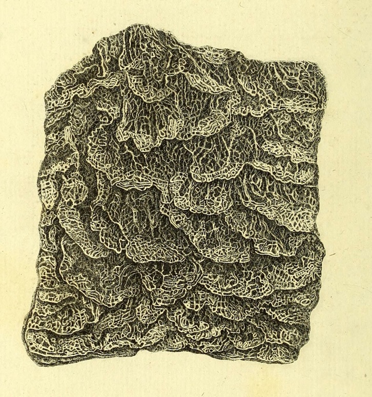
Johann Lieberkühn (1711-1756)
German physician, commemorated in crypts of Lieberkühn of the intestine.
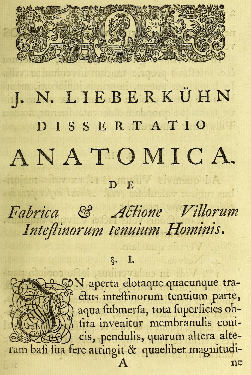
Lieberkühn began his academic career following his father's wish that he study theology in preparation for the ministry, but he also studied
natural sciences. After his father's death (and with permission from the king of Prussia to abandon his career in ministry),
Lieberkühn was able to devote his full attention to physical science and medicine. He obtained his medical degree from Leiden in 1739.
Lieberkühn invented improvements to his optical instruments. To examine blood vessels in microscopic detail
(capillaries had been first described by Malpighi in the previous century), Lieberkühn built
special-purpose microscopes, called "wonder-glasses" (Wundergläser) upon which a living animal, such as a frog, could
be fastened to observe the flow of blood. He invented the
Lieberkühn reflector to illuminate opaque specimens; this is a concave mirror surrounding the end of a microscope's
objective lens, to concentrate light directly upon the viewing area).
In his De fabrica et actione vollorum intestinorum tenuium hominis (1745), Lieberkühn describes, along with much else, the eponymous
intestinal "glands" for which he is remembered today. [The two images in this entry come from a 1782 edition available at the
Wellcome Collection.]
In his later career, Lieberkühn was noted for masterful preparation of durable preserved specimens, widely distributed for use in anatomical
demonstration.
For additional details of Lieberkühn's life and work, see Whonamedit
("a biographical dictionary of medical eponyms"). This entry includes further description of his inventions related to microscopy.
For a very brief bio, see Wikipedia.
A B C D
E F G H
I J K L
M N O P
Q R S T
U V W X
Y Z
TOP OF PAGE / ALPHABETICAL INDEX / CHRONOLOGICAL INDEX
James Lawrence Little (1836-1885)
American surgeon, commemorated in Little's area (also called Kiesselbach's area), a site in the anteroinferior nasal septum where an arterial plexus is a source for most nosebleeds.
Little's "vivid and clear" report of bleeding originating from the eponymous site was published as "A hitherto undescribed lesion as a cause of epistaxis, with four cases" (in The Hospital Gazette, v. 6, pp. 5-6) in 1879, several years prior to Kiesselbach's 1884 report on the same site. But it is the Kiesselbach eponym which seems to predominate, at least in sources of American origin. (The Little eponym seems to be used more commonly in the U.K., even though Little was an American.)
As noted by John J. Rainey in James
Lawrence Little, a forgotten pioneer (1952) AMA Arch. Otolaryngol., 55:451-452 (doi:10.1001/archotol.1952.00710010463007):
"[Little's] paper is not only the first description of the location in more than 90% of
all cases of nose bleeding, but it is the best ever written on the subject. Here is a great physician, with a talent for the clear, direct phrase, speaking to us across the years in simple, unadorned language...
"Kiesselbach, whose articles were published after Little's, at no time gave an exact description of the bleeding area as Little did, and his work, viewed from all angles, is decidedly inferior to Little's. Therefore, we should discontinue use of the term locus Kiesselbach and replace it with Little's area...
"It seems an ironic twist of fate that this man, whose name is not included in many medical dictionaries, should be neglected and almost forgotten in his own country, while his name is remembered in Great Britain by use of the term Little's area."
Little is mentioned very briefly in the Wikipedia entry on Kiesselbach's plexus; this is my only source for the dates of Little's birth and death.
A B C D
E F G H
I J K L
M N O P
Q R S T
U V W X
Y Z
TOP OF PAGE / ALPHABETICAL INDEX / CHRONOLOGICAL INDEX
Alexis Littre (1654-1726)
French physician and anatomist, commemorated in glands of Littre, small
periurethral mucous glands, mostly within the penis.
One publication relevant to the eponymous glands is available here (in French):
Alexis Littre: Description de l'urèthre
de l'homme, démontrée à l'Académie le troisième juillet 1700.
Mémoires de mathématique et de physique de l'Académie royale des sciences, 1700.
For a very brief biography listing a few more publications, see Whonamedit.
Wikipedia offers little more.
Additional English-language information about Littre has proven rather difficult to find. (This entire
webpage has been built while I sit at home surfing the Internet.)
A B C D
E F G H
I J K L
M N O P
Q R S T
U V W X
Y Z
TOP OF PAGE / ALPHABETICAL INDEX / CHRONOLOGICAL INDEX
Marcello Malpighi (1628-1694)
Italian scientist, commemorated in Malpighian corpuscles (i.e.,
renal corpuscles), the Malpighian layer of epidermis (i.e., stratum basale + stratum spinosum),
and Malpighian tubules (i.e., excretory organs of insects).
Malpighi is commonly designated as "the Father of Microscopic Anatomy." He is especially noted for describing
capillaries shortly after William Harvey had predicted
the necessity of just such invisibly small pores to connect arterial and venous circulation.
The life of Marcello Malpighi began at the dawning of scientific appreciation for optical instruments. At the time of Malpighi's
birth (1628), Galileo Galilei (1564 -1641) was still reporting wonders in the heavens discovered with his telescope. Malpighi's
contemporaries included pioneering microscopist Robert Hooke, whose work might have inspired Malpighi. Hooke
typically receives much more attention in introductory biology texts (for discovering "cells"), but Malpighi's contributions to anatomy were more considerable.
Malpighi was employed, at various times, on the medical faculties of Pisa, Messina, and Bologna. In the last few years of
his life he was private physician to Pope Innocent XII. Malpighi shared many of his observations, as did Hooke
and Leeuwenhoek, through letters to the Royal Society of London (founded in 1660); although Italian,
Malpighi became a Fellow of the Royal Society soon after (or an honorary member; consulted sources vary).
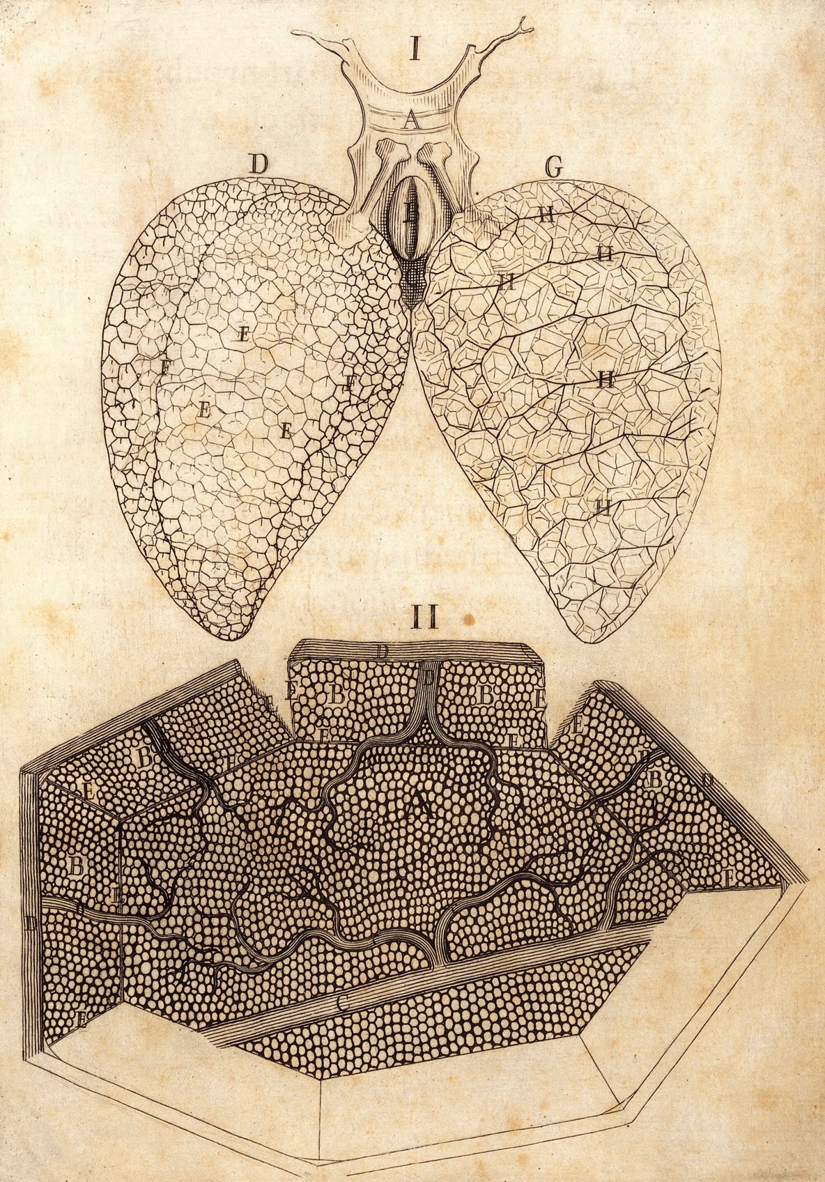 |
Malpighi's diagram of frog lung (top)
and alveolar capillaries (bottom),
from De pulmonibus... [ 2 ] |
The Encyclopedia Britannica's classic eleventh edition (1911; vol. 17, p. 497) offers a nice summary of Malpighi's research [ 1 ]:
"Malpighi was one of the first to apply the microscope to the study of animal and vegetable structure; and his discoveries were so
important that he may be considered to be the founder of microscopic anatomy...
"Although Harvey [i.e., William Harvey, who first described the circulation of blood in 1628] had correctly
inferred the existence of the capillary circulation, he had never seen it; it was reserved for Malpighi in 1661 (four years after Harvey's
death) to see for the first time the marvellous spectacle of the blood coursing through a network of small tubes on the surface of the
lung and of the distended urinary bladder of the frog..." [NOTE: Capillary circulation may be readily observed without
vivisection, albeit still with some inconvenience to the animal, by placing the tail of a living fish or the ear of a rabbit on the stage of a
microscope.]
"...[Malpighi's] discovery of the capillary circulation was given to the
world in the form of two letters De Pulmonibus; ...
these letters contained also the first account of the vesicular structure of the human lung, and they made a theory of respiration
for the first time possible... The achievement that comes next both in importance and in order of time was a demonstration of the plan
of structure of secreting glands; against the current opinion ... that the glandular structure was essentially that of a closed
vascular coil from which the secretion exuded, he maintained that the secretion was formed in terminal acini standing in open
communication with the ducts...
"The name of Malpighi is still associated with his discovery of the soft or mucous character of the lower stratum of the
epidermis [i.e., stratum basale + stratum spinosum], of the vascular coils in
the cortex of the kidney [i.e., renal corpuscles], and of the follicular bodies
in the spleen [i.e., splenic white pulp]. He was the first to attempt the finer anatomy
of the brain, and his descriptions of the distribution of grey matter and of the fibre-tracts in the cord, with their extensions
to the cerebrum and cerebellum, are distinguished by accuracy, but his microscopic study of the grey matter conducted him to the
opinion that it was of glandular structure and that it secreted the 'vital spirits.'"
Several of the above discoveries were published in De viscera structura exercitatio anatomica, 1666. A compelling account,
translated into English from Malpighi's own hand, is reproduced in "Completing the puzzle of blood circulation: the discovery of
capillaries," by M. Karamanou and G. Androutsos, in the Italian Journal of Anatomy and Embryology, v. 115, pp. 175-179
(2010), available
here (from ResearchGate) or here (from the National Library of
Medicine).
The above excerpts emphasize Malpighi's contributions to vertebrate histology. He also contributed more broadly to
microscopic anatomy in zoology and botany.
Early microscopists faced a challenge also shared by early telescopic astronomers -- establishing the reality of observed
phenomena, and then convincing others that their reports were not just artifacts. Early optical instruments certainly
did introduce artifactual appearances, notably diffraction fringes which can still mislead modern microscopists when condenser
and focus are not optimally adjusted [see Kohler illumination].
Tissue microscopists must contend not only with optical imperfections but also with
difficulties attending specimen preparation. Without fixation and staining, most biological tissues are relatively
uninformative. But microscopes and microscopic anatomy arose during the time of alchemy, when chemical knowledge was
quite limited. It took centuries to discover, through persistent trial and error, how to minimize artifacts such as
extraneous chemical precipitates and distortion of tissue and cell elements.
In 1683, in consideration of primitive histological protocols, ridicule was heaped on "those that flay dogs and cats, dry,
roast, bake, parboil, steep in vinegar, limewater, or aqua fortis livers, lungs, kidneys, calves' brains, or any other entrail,
and afterwards gaze on little particles of them through a microscope" [ 3 ].
(These were all methods for separating animal organs into component tissues. Even a century later, Bichat
was still subjecting his specimens "successively to desiccation, putrefaction, maceration, ebullition, stewing,
and to the action of the acids and the alkalis.") Renowned philosopher John Locke,
in spite of visiting Leeuwenhoek and seeing microscopic creatures for himself, remained unconvinced of the value of microscopy.
In his Essay Concerning Human Understanding (1689), Locke "insisted that if we had microscopes for eyes, the knowledge
we gained would be useless... God, in fact, has adapted our senses to our needs. Consequently, microscopical science
is 'lost labour' " [ 3 ].
Malpighi's observations were often conducted during vivisection rather than after chemical preservation. Nevertheless,
such work was repeatedly criticized by colleagues as having no medical value. Indeed, one of Malpighi's own students
"mounted a direct attack on his professor: 'It is our firm opinion that the anatomy of the exceedingly small, internal
conformation of the viscera, which has been extolled in these very times [i.e., by Malpighi] is of use to no physician' "
[3]. Indeed, "the immense impact of Malpighi's microscopic studies provoked envy and
criticism on the part of his contemporaries; a conflict that reached a boiling point in 1683 when his house was burnt and his manuscripts,
notes and laboratory equipment were destroyed completely" [ 4 ].
Toward the end of his life, Malpighi wrote his Opera Posthuma, which included an eloquent reply to his critics
[ 5 ]:
Nature ... in order to carry out the marvelous operations [that occur] in animals and plants has been pleased to construct
their organized bodies with a very large number of machines, which are of necessity made up of extremely minute parts so shaped
and situated as to form a marvelous organ, the structure and composition of which are usually invisible to the naked eye
without the aid of a microscope. ... Just as Nature deserves praise and admiration for making machines so small, so too
the physician who observes them to the best of his ability is worthy of praise, not blame, for he must also correct and repair
these machines as well as he can every time they get out of order.
But even in this posthumous work, Malpighi conceded that "microanatomy ... belonged to natural philosophy and not to
medicine" [ 3 ]. As a result of such criticism (and of such concessions), two centuries passed before microscopic anatomy
began to occupy a place in the standard medical curriculum.
Even in our own time, histology often receives less appreciation than other medical topics, perhaps because histology is
often presented to students more as a list of details to memorize than as a celebration "of extremely minute parts so shaped
and situated as to form a marvelous organ."
References cited above
- The classic 11th edition of The Encyclopedia Britannica (1911) is accessible through several online sources, including
here, at Wikisource.
- Marcello Malpighi, De pulmonibus observationes anatomicae (1661) [from Wellcome
Collection, Attribution 4.0 International (CC BY 4.0)].
- Quotations are from Bad Medicine: Doctors Doing Harm Since Hippocrates, by David Wootton, Oxford U.P. 2006 (accessible through GoogleBooks).
- M. Karamanou and G. Androutsos, Completing the puzzle of blood circulation: the discovery of capillaries, Italian Journal of Anatomy and Embryology, v. 115, pp. 175-179 (2010)
[available
here (from ResearchGate) or here (from the National Library of Medicine)] includes
not only Malpighi's own words, as mentioned above, but also additional fascinating information about his life.
- This translated excerpt from Opera Posthuma is reproduced in several sources.
Additional internet references
- Malpighi's Opera Omnia (1686),
at Real Jardín Botánico, Madrid.
- Malpighi's Opera Posthuma (1697), at the University of Utah's Digital Library.
- "Completing the puzzle of blood circulation: the discovery of capillaries," by M. Karamanou and G. Androutsos, in the Italian Journal of Anatomy and Embryology,
v. 115, pp. 175-179 (2010) [available
here (from ResearchGate) or here (from the National Library of Medicine)] includes
not only Malpighi's own words, as mentioned above, but also additional fascinating information about his life.
- A sampling of illustrations (including botany and invertebrate zoology) from Malpighi's Opera Omnia, as well as a nice essay:
here.
- An essay describing Malpighi's research on lung from the American Journal of Physiology-Lung Cellular and Molecular Physiology:
here.
- A nice biographical essay by Ghosh & Kumar, from the European Journal of Anatomy 22: 433-439 (2018):
here.
- Another biographical essay, this one by R.R. Reverón, from the International Journal of Morphology 29: 399-402 (2011):
here.
A B C D
E F G H
I J K L
M N O P
Q R S T
U V W X
Y Z
TOP OF PAGE / ALPHABETICAL INDEX / CHRONOLOGICAL INDEX
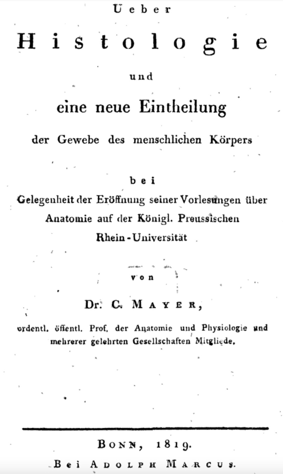 August Franz Joseph Karl Mayer (1787-1865)
German anatomist/physiologist. Mayer wrote on a wide range of topics (see publication list at
Wikipedia), often in support of abstract concepts in "Naturphilosophie" (more below).
We will here credit Karl Mayer for establishing the word "histology" (German, "histologie") as the name for a new science, in his 1819 book Ueber Histologie und eine
neue Eintheilung der Gewebe des menschlichen Körpers ["On histology and a new division of tissues of the human body"].
Prior to Bichat ("the Father of Histology"), histology did not yet exist as a distinct branch of anatomical science. And Bichat
himself did not provide a label for the discipline that he was founding. Some sources claim the word "histology" itself was coined by Craig; this is
presumably based on the "histology" entry in the Oxford English Dictionary which lists "Craig 1847, the doctrine of the organic tissues" as the first
use of the term in English. However, the equivalent term in German appeared decades earlier in the title for Mayer's brief tome, which reviewed Bichat's work.
Full text of Ueber Histologie... is available from GoogleBooks
One of Mayer's publications,
Die
Metamorphose der Monaden, which begins with extended quotes (in Latin) from Malpighi and Leeuwenhoek,
is intriguing from a histological perspective. The subject is the life of blood cells,
but for a modern reader Mayer's perspective in Naturphilosophie appears quite peculiar.
(Googling "metamorphosis of monads" takes one directly to philosophical works by
Leibnitz, on the nature of reality.) The following quotation is taken from N. Rüdinger (1885), at
Wikisource, quoting
Algemeine Deutsche Biographie.
"[Mayer's] research was broad in scope, extended to comparative anatomy [e.g., whale skin, fish brains], physiology, and anthropology
[e.g., Neanderthal fossils], and was all permeated by the speculative spirit that prevailed at the time... [A number of his writings]
contain sober, exact observations. But
besides the simple works on the umbilical vesicle, the cilia, the pharyngeal bursa, [and] the ganglia of the hypoglossal nerve, there are at the same time
others which address quite far-reaching questions, such as those on the law of gravity, reflex movement without spinal cord, yolk furrowing on the blood sphere,
and the like, works which contain results that are characteristic of the naturalist of that time. The natural processes were all too often exploited
only to gain apparent documentation for a priori philosophical ideas."
[Translation assisted by DeepL Translate and GoogleTranslate]
Brief biography, from Wikipedia.
(Not-quite-so-brief biography, from German-language Wikipedia.)
A B C D
E F G H
I J K L
M N O P
Q R S T
U V W X
Y Z
TOP OF PAGE / ALPHABETICAL INDEX / CHRONOLOGICAL INDEX
Johann Heinrich Meibom (1638-1700)
German anatomist and physician, commemorated in Meibomian glands
(sebaceous tarsal glands of the eyelid).
In the image at right (from Meibom's 1666 De
Vasis Palpebrarum Novis Epistola, "A New Letter about the Vessels of the Eyelid."), the skin surrounding the eye
is shown removed from the face. The eyelids are folded back, with eyelashes at F. The dots in a row along the
edge of each eyelid, at the base of the lashes, are pores for the Meibomian glands. These pores are paired with lines
(wiggly lines at E for the upper lid, straight lines at D for the lower lid) which indicate the positions
of the glands themselves.
(For a subsequent illustration of Meibom's glands, published a century later, see Zinn, below.)
At the University of Helmstedt, Meibom was not only professor of medicine but also professor of history
and poetry, a position which had previously been held by his
grandfather, with the same name. Meibom's work in history was published together with that of his grandfather in
Rerum germanicarum (1688).
Meibom also wrote latin poetry, as did his grandfather.
De Vasis Palpebrarum Novis Epistola may be viewed at
the Wellcome Collection.
Very brief biography at Whonamedit.com
Very brief biography at Wikipedia
A B C D
E F G H
I J K L
M N O P
Q R S T
U V W X
Y Z
TOP OF PAGE / ALPHABETICAL INDEX / CHRONOLOGICAL INDEX
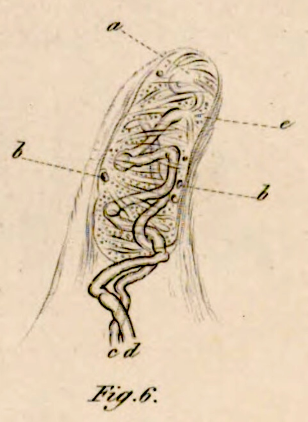 Georg Meissner (1829-1905)
German anatomist and physiologist, commemorated in Meissner's plexus (submucosal plexus of gastrointestinal tract) and
Meissner's corpuscles (tactile sensory receptors).
While studying medicine in Göttingen, Meissner collaborated with his professor Rudolf Wagner,
with whom he reported the discovery of the eponymous tactile corpuscles:
Wagner, R., & Meissner, G. (1852), "On the presence of previously unknown peculiar corpuscles (corpuscula tactus) in the sensory cusps of the human skin and on the end propagation of sensitive nerves" [Über das Vorhandensein bisher unbekannter eigentümlicher Tastkörperchen (corpuscula tactus) in den Gefühlswärzchen der menschlichen Haut und ü'ber die Endausbreitung sensitiver Nerven], Nachrichten von der Georg-Augusts-Universität und der Königlichen Gesellschaft der Wissenschaften zu Göttingen, Vol. 2, pp. 17-30.
The image to right, showing two nerve fibers entering and winding about
within a tactile corpuscle, is from Meissner's Beiträge zur Anatomie und Physiologie
der Haut (Leipzig 1853) [accessed at Bayerische Staatsbibliothek].
According to a fairly extensive entry for Meissner at Whonamedit.com, Meissner later reported the discovery under his own name,
in his dissertation, leading to conflict with Wagner over priority (with subsequent reconciliation).
Meissner's eponymous submucosal plexus is described in "About the nerves of the intestinal wall" [Über die Nerven der Darmwand],
Zeitschrift für rationelle Medizin, Neue Folge Vol. 8, pp. 364-366 (1857).
Meissner served as doctoral advisor for a notable student, Robert
Koch, the Nobel Prize-winning bacteriologist who is remembered in
"Koch's postulates." (For a modern perspective on Koch's postulates, see here.)
Additional biographical information can be found at Whonamedit.com and at
Wikipedia.
A B C D
E F G H
I J K L
M N O P
Q R S T
U V W X
Y Z
TOP OF PAGE / ALPHABETICAL INDEX / CHRONOLOGICAL INDEX
Friedrich Sigmund Merkel (1845-1919)
German anatomist, commemorated in Merkel discs and
Merkel cells of epidermis.
After receiving his doctorate, Merkel worked as prosector with Jakob Henle in Göttingen. He moved on to faculty positions in Rostok and in Königsberg before returning to Göttingen in 1885 to serve as Henle's successor.
Unlike receptor cells in organs of special sense, "the receptors in or beneath the surface
of the skin were generally named after those who first described them (e.g., Golgi tendon organs, Krause
end-bulbs, Meissner's corpuscles, Merkel discs, Pacinian
corpuscles, and Ruffini cylinders)" ["Receptor
Visionaries," by Nicholas Wade, Perception, 47: 833-850 (2018)].
In a multivolume textbook on human anatomy, Merkel introduced the now common practice in anatomical diagrams
of indicating arteries, veins, and nerves with the colors red, blue, and yellow
[per "Demystifying Merkel,"
JAMA Dermatol (2014) 150:814. doi:10.1001/jamadermatol.2014.225.]
Selected publication by Merkel:
- F. S. Merkel, "Über die Endigungen der sensiblen Nerven in der Haut der Wirbeltiere" [On sensory nerve terminations in the skin of vertebrates], Rostock, 1880.
Very brief biography at Wikipedia.
A B C D
E F G H
I J K L
M N O P
Q R S T
U V W X
Y Z
TOP OF PAGE / ALPHABETICAL INDEX / CHRONOLOGICAL INDEX
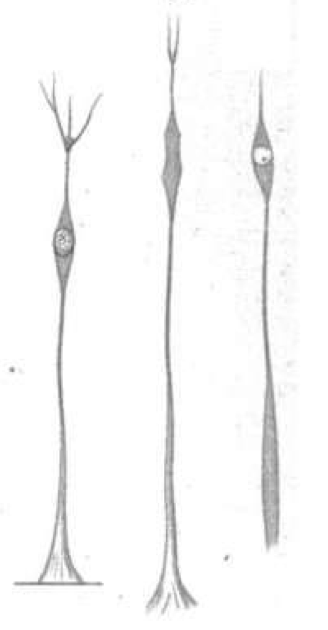 |
Müller's drawings of
retinal radial glial cells
Plate I, Fig. 6 in
Heinrich Müller's gesammelte und hinterlassene Schriften zur Anatomie und Physiologie des Auges, 1872.
Accessed
here. |
Heinrich Müller (1820-1864)
German anatomist, commemorated in Müller glial cells of the
retina.
A short account of Müller's glial-cell observations can be found in section 3.1 of the following report:
Chvátal, A. & Verkhratsky, A. (2018). An Early History of Neuroglial Research: Personalities. Neuroglia 1(1):245-257. https://doi.org/10.3390/neuroglia1010016
Müller is especially noted for his clever demonstration that vision is initiated with rods and cones
at the back of the retina, using precise geometric calculations based on entoptic observation of retinal
blood vessels (i.e., the Purkinje tree,
first reported by Jan Purkinje in 1819):
Müller H. (1855) Ueber die entoptische Wahrnehmung der Netzhautgefässe, insbesondere als Beweismittel für die Lichtperception durch die nach hinten gelegenen Netzhautelemente [On the entoptic perception of retinal vessels, in particular as evidence for light perception through the backwards-lying retinal elements]. Verhandlungen. Physikalisch-Medizinische Gesellschaft in Würzburg 5:411-447.
Translated excerpts from this report can be found in
J.S. Warner, et al. (2013) Heinrich Müller (1820-1864) and the entoptic discovery of the site in the retina where vision is initiated," Journal of the History of the Neurosciences 31:64-90 DOI: 10.1080/0964704X.2021.1959165.
(Credit for discovering which retinal layer contains photoreceptor cells is shared with Carl Bergmann, who
followed a different line of reasoning, that the fovea where vision is most acute contains only rod and cone cells.)
Brief biography at Portraits of European Neuroscientists
Brief biography at Wikipedia
Brief biography at Whonamedit
A B C D
E F G H
I J K L
M N O P
Q R S T
U V W X
Y Z
TOP OF PAGE / ALPHABETICAL INDEX / CHRONOLOGICAL INDEX
Martin Naboth (1675-1721)
German physician and anatomist, commemorated in Nabothian cysts (mucus retention cysts) of the
cervix.
Naboth's observations were reported (in Latin, of course) in De Sterilitate Mulierum (1707). A facsimile
of this volume may be viewed here, at the
Bayerische Staatsbibliothek (Bavarian State Library).
Naboth mistakenly believed that he had discovered a site where eggs were stored. And although Naboth received the eponym,
these cysts had been previously described in 1681 by French surgeon Guillaume des Noues. (Des Noues, by the way,
collaborated for a time with
Gaetano Giulio Zumbo, who has been described as the founder of anatomic wax modeling.)
Brief biography at "Medical Terminology Daily."
A B C D
E F G H
I J K L
M N O P
Q R S T
U V W X
Y Z
TOP OF PAGE / ALPHABETICAL INDEX / CHRONOLOGICAL INDEX
Franz Nissl (1860-1919)
German neuropathologist / neuroanatomist commemorated in the Nissl stain
and the associated Nissl bodies.
Nissl studied medicine at the University of Munich, where he based a prize-winning essay on fixation and
staining techniques that he had developed. His doctoral dissertation, "Results
and experiences in examining the pathological changes in the nerve cells in the cerebral cortex" [Resultate und Erfahrungen bei
der Untersuchung der pathologischen Veränderungen der Nervenzellen in der Grosshirnrinde, University of Munich, 1885] was written
on the same topic as this essay. For the remainder of his career, Nissl continued research into the
histopathological correlates of mental disease. Among his colleagues and collaborators were Korbinian
Brodmann (of "Brodmann's areas" fame) and especially Alois
Alzheimer (of "Alzheimer disease" fame).
The Nissl stain is an historically important method of accentuating nerve cell bodies. This method
greatly facilitated the mapping of central nervous tissue, notably by Brodmann
and by Ramón y Cajal. Using this stain, Nissl was able to describe cellular changes associated with
retrograde degeneration. This in turn became the basis of a powerful tool for mapping pathways
within the brain, which Nissl himself used to study connections between thalamus and cerebral cortex.
Among Nissl's works are:
"About a new method of investigation of the central organ to determine the localization of nerve cells" [Ueber eine neue Untersuchungsmethode des Centralorgans zur Feststellung der
Localisation der Nervenzellen], Centralblatt für Nervenheilkunde und Psychiatrie, Coblenz: W. Groos, 1894, 13: 507-508 (available at The Wellcome Collection).
As editor, along with Alzheimer:
"Histological and histopathological works on the cerebral cortex: with special
reference to the pathological anatomy of mental diseases" [Histologische
und histopathologische Arbeiten über die Grosshirnrinde: mit besonderer Berücksichtigung der pathologischen
Anatomie der Geisteskrankheiten], Jena: Fischer, 1904-1913 (available at The Wellcome Collection).
Additional links:
Franz Nissl (1860-1919) Neuropathologist, Journal of the American Medical Association, 205: 460-461 (doi:10.1001/jama.1968.03140320154019) offers some detail on Nissl's research. [Also see Alois Alzheimer (1864-1915) Neurohistopathologist, Journal of the American Medical Association, 208: 1017-1018 (doi:10.1001/jama.1969.03160060087015).]
The Nissl entry at Whonamedit.com includes a substantial bibliography, while Nissl's relationship with Alzheimer is described in greater detail in that website's Alzheimer entry.
Franz Nissl, at Wikipedia.
Nissl method, at Wikipedia.
Nissl bodies, at Wikipedia.
A B C D
E F G H
I J K L
M N O P
Q R S T
U V W X
Y Z
TOP OF PAGE / ALPHABETICAL INDEX / CHRONOLOGICAL INDEX
Jean-Pierre Nuel (1847-1920)
Luxembourgian-Belgian ophthalmologist, commemorated in "Nuel's space" within the
organ of Corti of the inner ear.
Nuel reported his research on the mammalian cochlea in both German (1872) and French (1878), offering
additional detail and correction to prior reports by Boettcher and Deiters.
For more on Nuel's space, see
Cochlear Explorers - Part VIII
-- Space of Nuel. For Nuel's space and additional eponyms associated with the inner ear, see
J. Schacht & J.E. Hawkins,
"A Cell by Any Other Name: Cochlear Eponyms" (Audiology & Neuro Otology 2004, vol. 9, pp. 317-327, DOI: 10.1159/000081311).
Brief biography at Wikipedia.
A B C D
E F G H
I J K L
M N O P
Q R S T
U V W X
Y Z
TOP OF PAGE / ALPHABETICAL INDEX / CHRONOLOGICAL INDEX
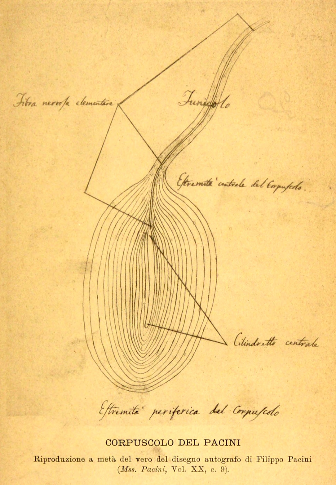 |
| Pacini's drawing, included in Aurelio Bianchi's "Report and catalog of Filippo Pacini's manuscripts in the National
Central Library of Florence" [Relazione e catalogo dei manoscritti di Filippo Pacini esistenti nella R. Biblioteca nazionale centrale di
Firenze], 1889. Image accessed at
The Wellcome Collection.] |
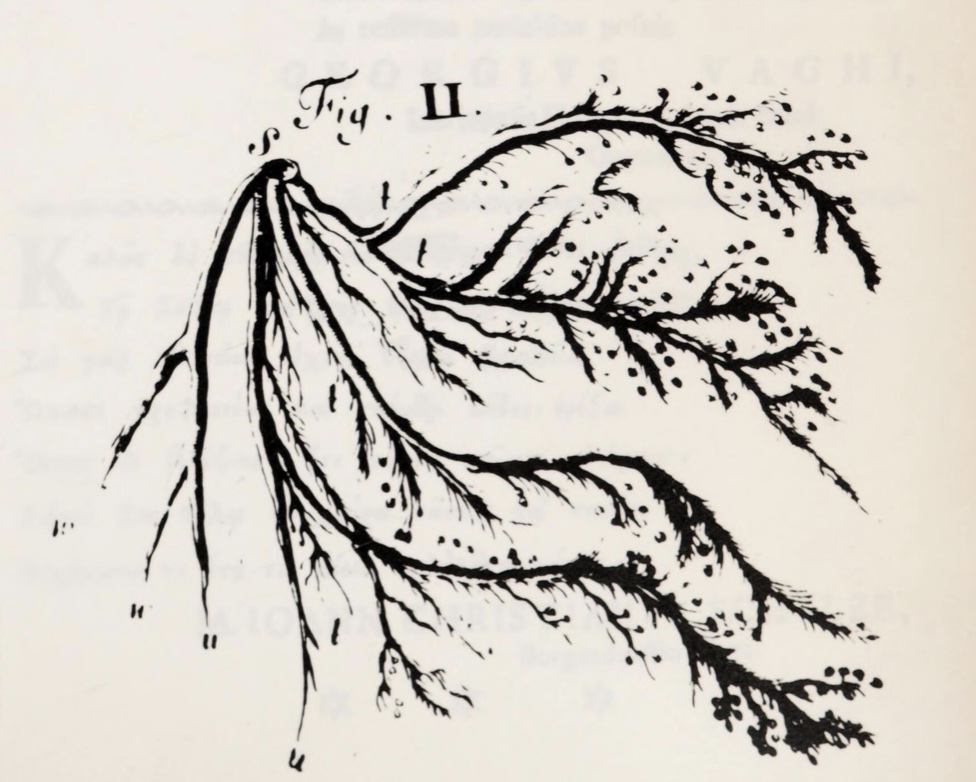 |
Vater's 1741 drawing showing "Papillae Nerveae, attached in large quantities to the digital nerves" (quote from
Bentivoglio & Pacini 1995, below). [Image accessed at The
Wellcome Collection.]
|
Filippo Pacini (1812-1883)
Italian anatomist, commemorated in Pacinian corpuscles of skin.
As a 19-year-old medical student in 1831, Pacini noticed the eponymous corpuscles during dissection and reported his
observation in 1835. He was not aware that the corpuscles had been previously observed and reported nearly a century
earlier, by Abraham Vater (who is himself commemorated in the ampulla of Vater,
of the hepatopancreatic duct).
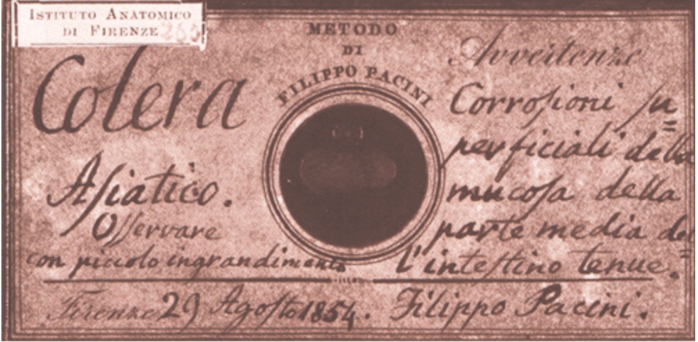 |
| "This microscope slide, prepared by Pacini in 1854, was clearly identified as containing the cholera
bacillus." (Image and caption from
Wikimedia Commons.) |
In 1854, during a widespread cholera epidemic, Pacini discovered and reported on the causative agent for the disease. 30 years later,
the cholera vibrio was rediscovered and reported by Robert Koch. Both discoveries were initially ignored or discounted by the medical establishment
of the time, in favor of the prevailing miasmic theory of contagion. Koch's reputation eclipsed that of Pacini, so for many years
Koch was credited with the discovery. Nevertheless, Pacini's priority was eventually recognized (see
here), and in 1965 Pacini was finally
and officially acknowledged as author of both genus and species of Vibrio cholerae Pacini 1854, by the
Judicial Commission of the International
Committee on Bacteriological Nomenclature. This story is nicely told in
"Who First Discovered Vibrio cholera,"
at the John Snow website. (John Snow was the famed London doctor and pioneering epidemiologist
who during the same 1854 epidemic determined that cholera was a waterborn disease, infamously spread by
"the Broad Street pump.")
A readable, and much more complete, account of Pacini's research is available
here, from M. Bentivoglio and P. Pacini, "Flippo Pacini: a determined
observer," Brain Research Bulletin Vol. 38, pp. 161-165 (1995). (For more, see "
The greatest steps towards the discovery of Vibrio cholerae.")
Brief biographies, each including summary accounts of Pacini's discoveries both of the tactile
corpuscles and of the cholera vibrio, are offered by Wikipedia and by
Whonamedit.com.
A B C D
E F G H
I J K L
M N O P
Q R S T
U V W X
Y Z
TOP OF PAGE / ALPHABETICAL INDEX / CHRONOLOGICAL INDEX
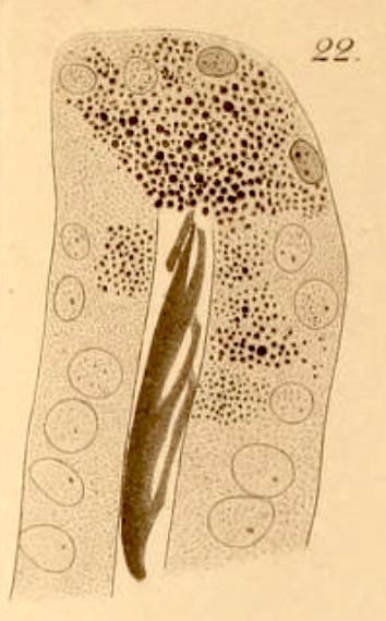 Josef Paneth (1857-1890)
Austrian physiologist, commemorated in Paneth cells
The intestinal crypt cells now known as Paneth cells were first described by
Gustav Schwalbe (Austrian anatomist, 1844-1916) in
Archiv für mikroskopische Anatomie, vol. 8, pp. 92-140 (1872).
Paneth subsequently recognized their secretory function, which he reported sixteen years later in the same journal, vol. 31, pp. 113-191 (1888),
"Ueber die secernirenden Zellen des Dünndarm-Epithels" ("About the secreting
cells of the epithelium of the small intestine"). As suggested by the journal title Archiv für mikroskopische Anatomie,
by the late 1800s microscopic anatomy had become a well-developed science.
The caption for the image at right (accessed
here, at the
Biodiversity Heritage Library) reads:
Crypt of Lieberkühn of the mouse. Fixation in picric acid, staining with saffranin according
to Pfitzner. Granule cells [i.e., Paneth cells] with granules of different sizes, those most plump and filled with the largest
granules in the fundus; secreted mass confluent in the fundus of the crypt.
"Lieberkühn'sche
Krypte der Maus. Härtung in pikrinsäure, Färbung mit Saffranin nach Pfitzner. Körnchenzellen mit verschieden grossen Körnchen,
die am prallsten und mit den grössten körnchen gefüllten im Fundus; Secretmasse confluirt
im Fundus der Krypte."
The full plate [here] includes images of crypts from dog and
human; the full article [here] includes as well extensive
description and illustration of goblet cell secretion in a newt.
Online biographies of Paneth commonly highlight his relationships with Sigmund
Freud and Friedrich Nietsche.
Brief bio at Wikipedia.
A B C D
E F G H
I J K L
M N O P
Q R S T
U V W X
Y Z
TOP OF PAGE / ALPHABETICAL INDEX / CHRONOLOGICAL INDEX
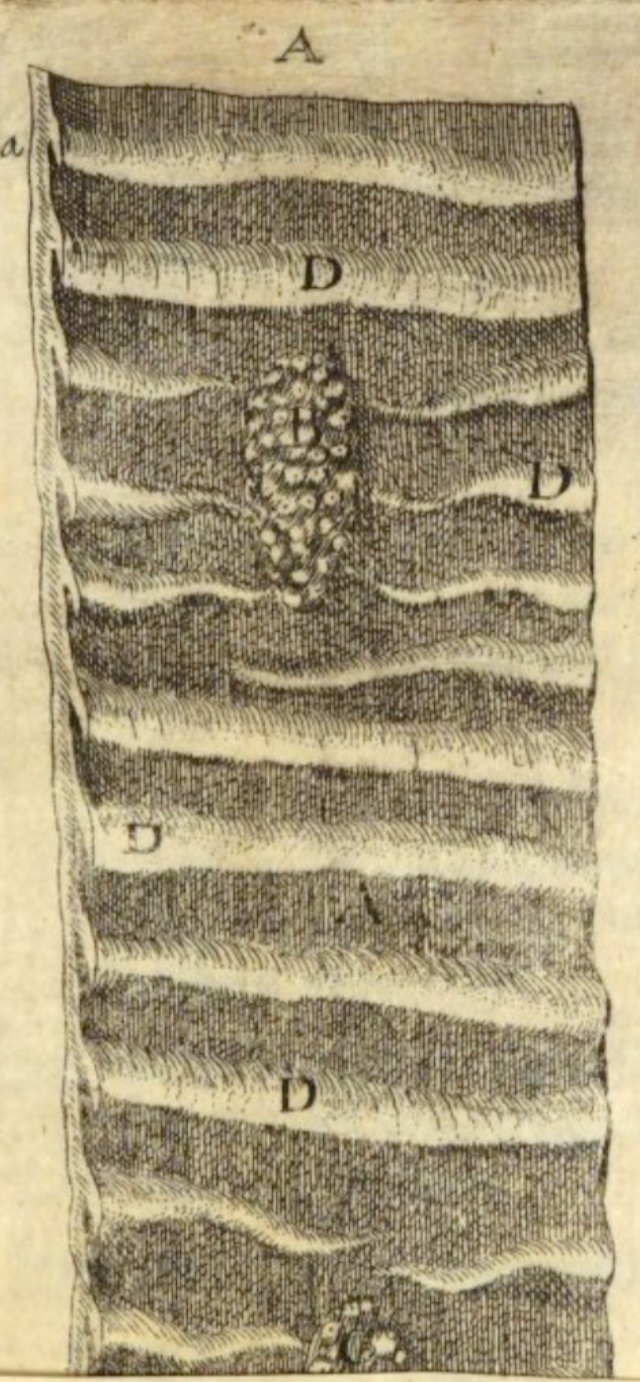
Johann Conrad Peyer (1653-1712)
Commemorated in Peyer's patches of the small intestine.
Peyer was born and died in Schaffhausen, Switzerland, but studied medicine in Paris and in Basel.
He was a pupil of Johann Jacob Wepfer (1620-1695, founder of the "Schaffhouse School" of anatomy and physiology,
in Schaffhausen); he worked with Webper and Johann Conrad von Brunner on gastrointestinal anatomy.
Peyer described the eponymous lymphoid patches as plexus glandularum (labelled B in the image at right)
in his 1682 publication Exercitatio anatomico-medica de Glandulis intestinorum ("Anatomical-Medical Study of
the Intestinal Glands"), available at the
Munich DigitiZation Center.
See [here] for a fascinating essay on the 17th century "Schaffhouse School"
of anatomy and physiology; Peyer is mentioned on p. 515.
The Anatomical and Physiological Approach in Swiss Medicine during the 17th Century, by Heinrich Buess, Bulletin of
the History of Medicine, (Vol. 27, pp. 512-520, 1953), The Johns Hopkins University Press.
Listing of biographical details, here from Rice Univ.
Brief bio, from Wikipedia
More on Peyer's patches, from Wikipedia.
A B C D
E F G H
I J K L
M N O P
Q R S T
U V W X
Y Z
TOP OF PAGE / ALPHABETICAL INDEX / CHRONOLOGICAL INDEX
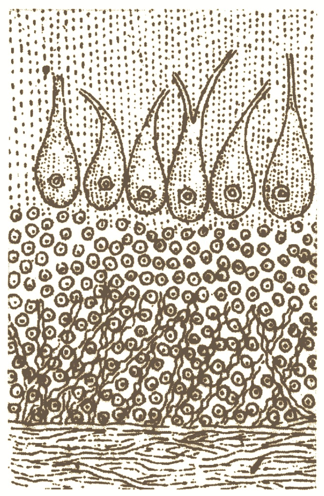 Jan (or Johann) Evangelista Purkinje (1787-1869)
Bohemian anatomist, commemorated in Purkinje cells of
cerebellum and Purkinje fibers of heart, also the
Purkinje shift in color vision and
the Purkinje tree of retinal vasculature.
(Heinrich Müller later used entoptic observations of the Purkinje tree to infer
that rods and cones were the site for initiation of visual perception.)
Purkinje earned his medical degree in Prague in 1818, where he then served for a few years as prosector in anatomy. In 1823 he was
appointed as Professor of Physiology and Pathology at the Royal Prussian University of Wroclaw
(=Breslau, in what is now Poland), where he had to wait nine years to be granted his first microscope.
In 1850 he returned to Prague for a chair in Physiology of the Prague Medical Faculty.
During a time when Prussia (and the German language) dominated the areas now known
as the Czech Republic and Poland, Purkinje promoted Czech independence as well as the Czech and Polish languages.
Purkinje's research engaged with many areas of science, from heart function and brain
anatomy to pharmacology (he tested drugs on himself, a not-uncommon practice
at the time), optics, vision (his work on color perception caught the attention of poet/scientist
Johann Wolfgang von Goethe), and the classification
of fingerprints. He advanced the development of histological techniques;
some sources attribute to Purkinje the development of the first practical
microtome for use in tissue sectioning.
He elucidated the electrical system of the heart, describing the conducting fibers which bear his name. In 1837, at a time before
nervous tissue was well understood (long before Cajal had clarified the Neuron Doctrine, even before Cell
Theory had become familiar), Purkinje described "ganglionic corpuscles" (i.e., nerve cell bodies) in many regions of the brain,
including the cerebellar cortex where Purkinje cells form a distinctive layer:
Ueber die gangliösen Körperchen in verschiedenen Theilen des Gehirns ["On the ganglionic corpuscles in various parts of the
brain"] (1837), Ber. Naturf. Prague, p. 137.
Biography by Minai Mandana, Embryo Project Encyclopedia (2014-06-05), ISSN: 1940-5030 https://hdl.handle.net/10776/7898, Arizona State University.
Biography from the Texas Heart
Institute Journal, Feb. 2018, Vol. 45, No. 1
Biography in Advances in Physiological Education, with
extensive description of Purkinje's research results in several areas.
Jan Voogd, The Purkinje Cell (Ch. 6, pp. 37-43, in Neurological Eponyms, P. J. Koehler et al., eds., Oxford
University Press, 2000), available through Google Books.
(Use the "search" function at GoogleBooks to find this chapter.)
A B C D
E F G H
I J K L
M N O P
Q R S T
U V W X
Y Z
TOP OF PAGE / ALPHABETICAL INDEX / CHRONOLOGICAL INDEX
Santiago Ramón y Cajal (1852-1934): See Cajal, above.
A B C D
E F G H
I J K L
M N O P
Q R S T
U V W X
Y Z
TOP OF PAGE / ALPHABETICAL INDEX / CHRONOLOGICAL INDEX
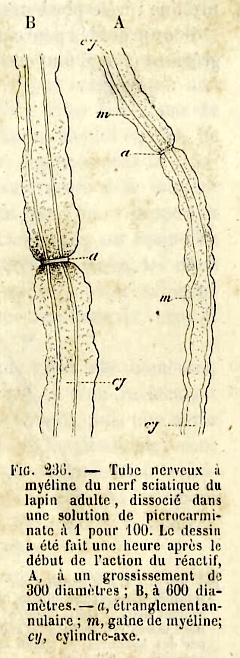 Louis Antoine Ranvier (1835-1922)
French physician, pathologist, anatomist and histologist, commemorated in nodes of Ranvier.
The image at right -- one of Ranvier's eponymous nodes, here labelled "étranglement
annulaire" ("annular constriction") -- was taken from Ranvier's Traité technique d'histologie (1875), available at the
Wellcome Collection. This 820 page treatise provides an illustrated
overview (in French) of histological knowledge in the second half of the nineteenth century.
Ranvier's research in many aspects of histology emphasized the radical (for his time) concept that
histological study could elucidate organ physiology. For example:
"[Ranvier's] description of nerve fibre nodes was made in a search for how nutrients were continuously exchanged with the blood for
nerve cell function... Physiology had demonstrated a loss of motor nerve function by interruption
of blood flow and a return to function by perfusion of oxygenated blood... The question was then clear to Ranvier: what is
the path for oxygen between oxygenated blood and nerve fibers? For Ranvier, the continuous and impermeable myelin sheath
of nerve fibres prevented exchange of fluids and thereby nutrition. He demonstrated the point histologically showing that
... picrocarminate could penetrate fibres, at localized sites identified as interruptions of the myelin sheath..."
The above quote is taken from an excellent, illustrated overview of Ranvier's research by Jean Gaël Barbara,
available here [from
IBRO, the International Brain Research Organization].
Very brief summaries of Ranvier's career can be found here
(from Wikipedia) and here (from Nature, 1935), but these provide very minimal information
about Ranvier's research.
A B C D
E F G H
I J K L
M N O P
Q R S T
U V W X
Y Z
TOP OF PAGE / ALPHABETICAL INDEX / CHRONOLOGICAL INDEX
Ernst Reissner (1824-1878)
German anatomist, commemorated in Reissner's membrane (the vestibular membrane) of the cochlea.
Reporting in "Zur Kentniss der Schnecke im Gehörorgan der Säugethiere und des Menschen" [Toward understanding the cochlea in
the auditory organs of mammals and humans], (Arch Anat Physiol Wiss Med (1854), pp 420-427), Reissner effectively discovered the endolymphatic
passageway, now called the scala media or cochlear duct, by describing the eponymous
membrane which separates it from the scala vestibuli.
For more on Reissner's membrane and additional eponymous structures associated with the inner ear, see
J. Schacht & J.E. Hawkins,
"A Cell by Any Other Name: Cochlear Eponyms" (Audiology & Neuro Otology 2004, vol. 9, pp. 317-327, DOI: 10.1159/000081311).
Brief biography at Wikipedia.
A B C D
E F G H
I J K L
M N O P
Q R S T
U V W X
Y Z
TOP OF PAGE / ALPHABETICAL INDEX / CHRONOLOGICAL INDEX
Charles-Philippe Robin (1821-1885)
French anatomist and biologist, commemorated in Virchow-Robin space (perivascular space in the brain, essentially extending
subarachnoid space along blood vessels).
Brief biography at Wikipedia.
More extensive biography, at Whonamedit.com.
A B C D
E F G H
I J K L
M N O P
Q R S T
U V W X
Y Z
TOP OF PAGE / ALPHABETICAL INDEX / CHRONOLOGICAL INDEX
Friedrich-Christian Rosenthal (1780-1829)
German surgeon and anatomist, commemorated in Rosenthal's canal (housing the
spiral ganglion of the cochlea) and the basal vein of Rosenthal.
Pursuing various opportunities, Rosenthal relocated several times within Germany and Austria,
eventually accepting a position as full professor and museum director at a university
in Greifswald, the town of his birth and education. Details of this life, including the
several anatomists with whom he worked, are recounted (in German) in
Allgemeine Deutsche Biographie.
(This 1889 entry may be easily translated by copy-and-pasting into GoogleTranslate
or DeepL.)
Rosenthal described the structure now known as Rosenthal's canal in a report on the structure of the modiolus in
the human ear (Über den Bau der Spindel im menschlichen Ohr, 1823). He described the vena
basilaris Rosenthalii in a report on branches of the great vein of Galen (De intimis cerebri venis
seu de venae magnae Galeni ramis, 1824). This vein was given its eponymous designation by Joseph Hyrtl, in his 1846
"Textbook of human anatomy" (Lehrbuch der Anatomie des Menschen). During his relatively short career,
Rosenthal also published extensive works on the anatomy of whales, seals, and sea-lions. But much of his work
remained incomplete, interrupted by his untimely death from tuberculosis at age 49.
Additional resources for Rosenthal:
Brief biography from
Hearing: Health and Technology Matters.
Very brief biography at Wikipedia.
A B C D
E F G H
I J K L
M N O P
Q R S T
U V W X
Y Z
TOP OF PAGE / ALPHABETICAL INDEX / CHRONOLOGICAL INDEX
Charles Marie Benjamin Rouget (1824-1904)
French physiologist, commemorated in Rouget cells, cells associated with capillaries
that are now known as pericytes.
Rouget studied medicine at hospitals in Paris and became professor of physiology at the University of Montpellier and at the Muséum
d'Histoire Naturelle in Paris. He reported the eponymous cells in 1874 in Note sur le developpement de la tunique
contractile des vaisseaux (Compt. Rend. Acad. Sci., vol. 59, pp. 559-562).
The history of pericyte research is briefly recounted in the introduction to
Morphology and properties of pericytes by
P. Dore-Duffy and K. Cleary, Methods in Molecular Biology, vol 686, pp. 49-68 (2011).
[NOTE: This link appears to open the wrong page, but scrolling down leads to the full text of the 2011 pericyte review.
The "proper" link for this review,
doi.org/10.1007/978-1-60761-938-3_2, opens an abstract only.
(For a more thorough account of historical understanding of capillaries, see
"The history of the capillary wall:
doctors, discoveries, and debates," by C. Hwa and W.C. Aird, in
Am J Physiol Heart Circ Physiol 293: H2667-H2679 (2007),
doi:10.1152/ajpheart.00704.)
Additional resources:
A B C D
E F G H
I J K L
M N O P
Q R S T
U V W X
Y Z
TOP OF PAGE / ALPHABETICAL INDEX / CHRONOLOGICAL INDEX
Angelo Ruffini (1864-1929)
Italian anatomist, commemorated in Ruffini corpuscles (Ruffini nerve endings).
"Among the microscopic structures that were isolated and described after the cell
doctrine had been enunciated were specialized sensory cells, called receptors... Those located in well-defined sense organs
were named on the basis of their morphology (rods, cones, hair cells, etc.), whereas the receptors in or beneath the surface
of the skin were generally named after those who first described them (e.g., Golgi tendon organs, Krause
end-bulbs, Meissner's corpuscles, Merkel discs, Pacinian
corpuscles, and Ruffini cylinders)" [quote from "Receptor
Visionaries," by Nicholas Wade, Perception, 47: 833-850 (2018)].
Selected publications by Ruffini:
- A. Ruffini, "Di una particolare reticella nervosa e di alcuni corpuscoli del Pacini che si trovano in connessione cogli organi muscolo-tendinei del gatto" [Of a particular nervous net and some Pacini corpuscles that are found in connection with the muscle-tendon organs of the cat], Rendiconti. Classe di scienze fisiche, matematiche e naturali, 5th ser., vol. 7 no. 1, 442-446.
- A. Ruffini, 1896. Sulla fine anatomia dei fusi neuro-muscuolare del gatto e sul loro significato fisiologico. Monitore Zoologico Italiano 7, 49-52.
- A. Ruffini, 1898. On the minute anatomy of the neuromuscular spindles of the cat, and on their physiological significance. J. Physiol. 23, 191-208
Ruffini corpuscles at Wikipedia.
Very brief biography at Wikipedia.
A B C D
E F G H
I J K L
M N O P
Q R S T
U V W X
Y Z
TOP OF PAGE / ALPHABETICAL INDEX / CHRONOLOGICAL INDEX
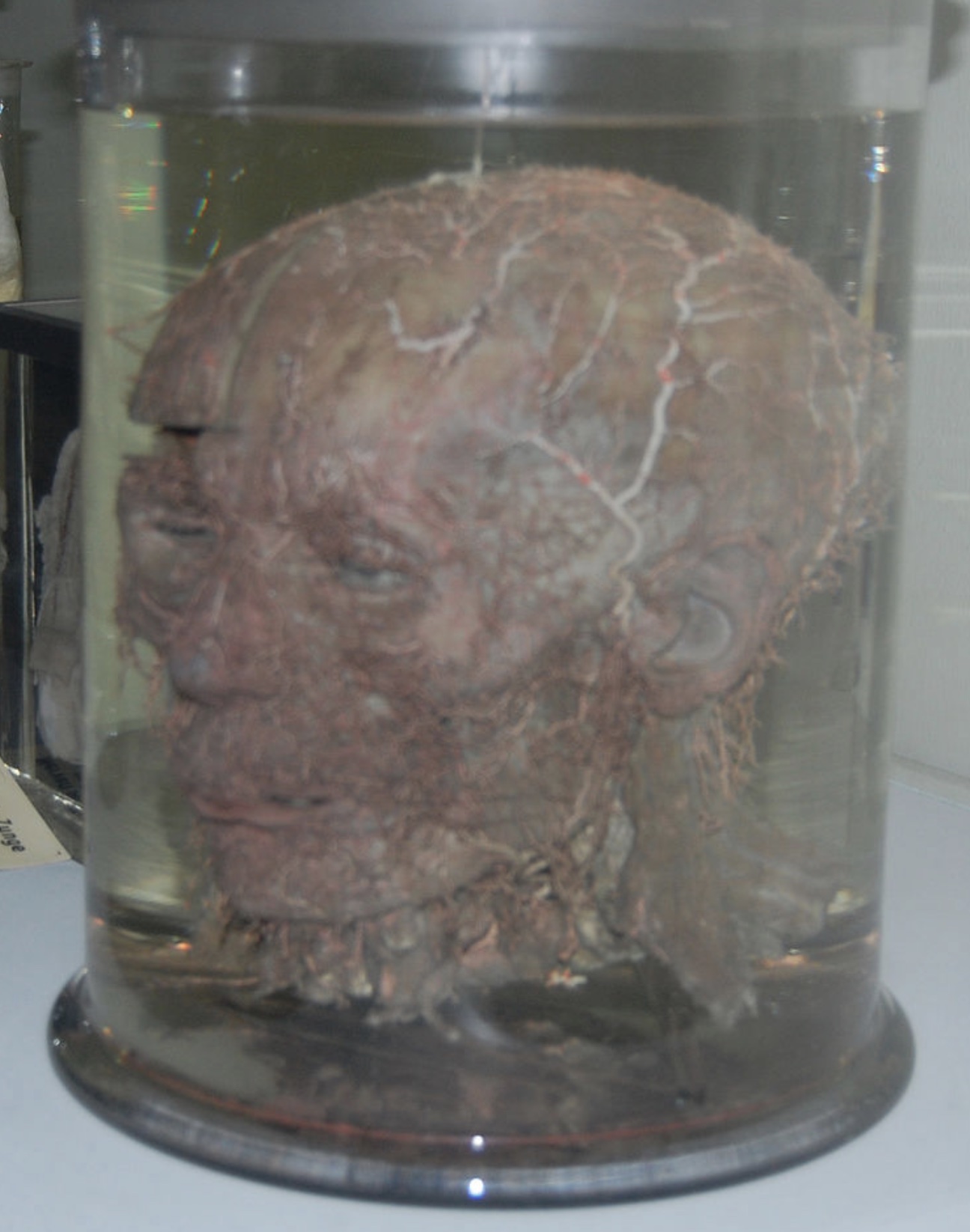 |
Image from Wikimedia Commons:
"Skull with meticulously separated arteries prepared by Frederich Schlemm. Displayed in the Center for Anatomy of Charite (University of Berlin)." |
Friedrich Schlemm (1795-1858)
German anatomist, commemorated in the canal of Schlemm of the eye.
Schlemm advanced to full professor at the University of Berlin, earning fame for his superb dissection technique. Some of his preparations
have been preserved, such as that shown in the image at right. His best-known published work is an 1830 desciption of the superficial arteries of
the head, Arteriarum capitis superficialum icon nova. (On the page
opened by this link, look for "Download / PDF." Schlemm's illustrations at the end of this short monograph will reward the download.)
Schlemm's discovery of the eponymous canal is briefly described in "Eyeing
the Eye" (an article by Nicole Davis, at The Jackson Laboratory, in Search Magazine, 19 Dec. 2014).
Unfortunately this article does not cite a source for the plausible story that Schlemm found the canal because it had become "engorged with blood"
in the corpse of a man who had
hanged himself.
A 2008 article in the Annals of Anatomy (Anatomischer Anzeiger) reports that
in 1816 a much younger Schlemm had been sentenced to a month in prison, after being apprehended for disinterring the body of deceased woman.
"Body-snatching" was not unusual in the early nineteenth century, as a method for acquiring cadavers for medical dissection (in this case, for studying
effects of rickets on bones at the Anatomico-Surgical Institute in Braunschweig). (For more on "the resurrectionists," see
Art macabre: Resurrectionists and anatomists,
R. McGee, 2001, ANZ Journal of Surgery, vol. 71, pp.377-380.)
The entry for Schlemm at Wikipedia offers little more than a summary of the article
cited above, from the German Annals of Anatomy.
An entry (in German) at Allgemeine Deutsche Biographie offers some additional
but dated (1890) detail (this note can be easily copy-and-pasted into DeepL Translator or
Google Translate).
A B C D
E F G H
I J K L
M N O P
Q R S T
U V W X
Y Z
TOP OF PAGE / ALPHABETICAL INDEX / CHRONOLOGICAL INDEX
Henry D. Schmidt (1823-1888)
American anatomist, surgeon and pathologist, commemorated by Schmidt-Lanterman clefts in myelin.
Schmidt was born in Germany, emigrated to Pennsylvania at age 25, and graduated in medicine from the University of Pennsylvania in 1858. From there he went to the Medical College of Alabama and then to the New Orleans School of Medicine. During the Civil War, Schmidt served the Confederacy as field surgeon; he continued after the war as pathologist with Charity Hospital in New Orleans. He is reported to have opposed Koch's discovery of tuberculosis bacilli, arguing that they were merely artifacts.
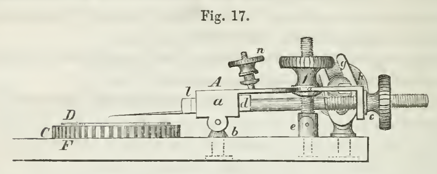 |
Schmidt's micromanipulator,
image accessed at Wikimedia |
Schmidt's discovery of the eponymous myelin clefts was reported in "On the Construction of the Dark or Double-bordered Nerve Fibre" in The Monthly Microscopical Journal (vol. 11:200-221) in 1874, the same year as Lanterman independently reported the same structure. His 1859 report "On the Minute Structure of the Hepatic Lobules" in The American Journal of the Medical Sciences (vol. 37, p. 13-39) includes a detailed description of his multi-tool micro-manipulation device, designed to enable "the slightest motion of microscopic needles, knives, or scissors, in different directions."
In 1884, The New Orleans Medical and Surgical Journal (vol. 12, pp. 325-333) published a magnificently florid ovation / eulogium on Schmidt, upon completion of the "New Pathological Department" at Charity Hospital in New Orleans. (This hospital gained notoriety following Hurricane Katrina in 2005.)
My principal starting point for this entry was the Polish Wikipedia with its included links. Also see American Medical Biographies.
A B C D
E F G H
I J K L
M N O P
Q R S T
U V W X
Y Z
TOP OF PAGE / ALPHABETICAL INDEX / CHRONOLOGICAL INDEX
Theodor Schwann (1810-1882)
German physician, commemorated in Schwann cells, also celebrated as one of the founders
of Cell Theory.
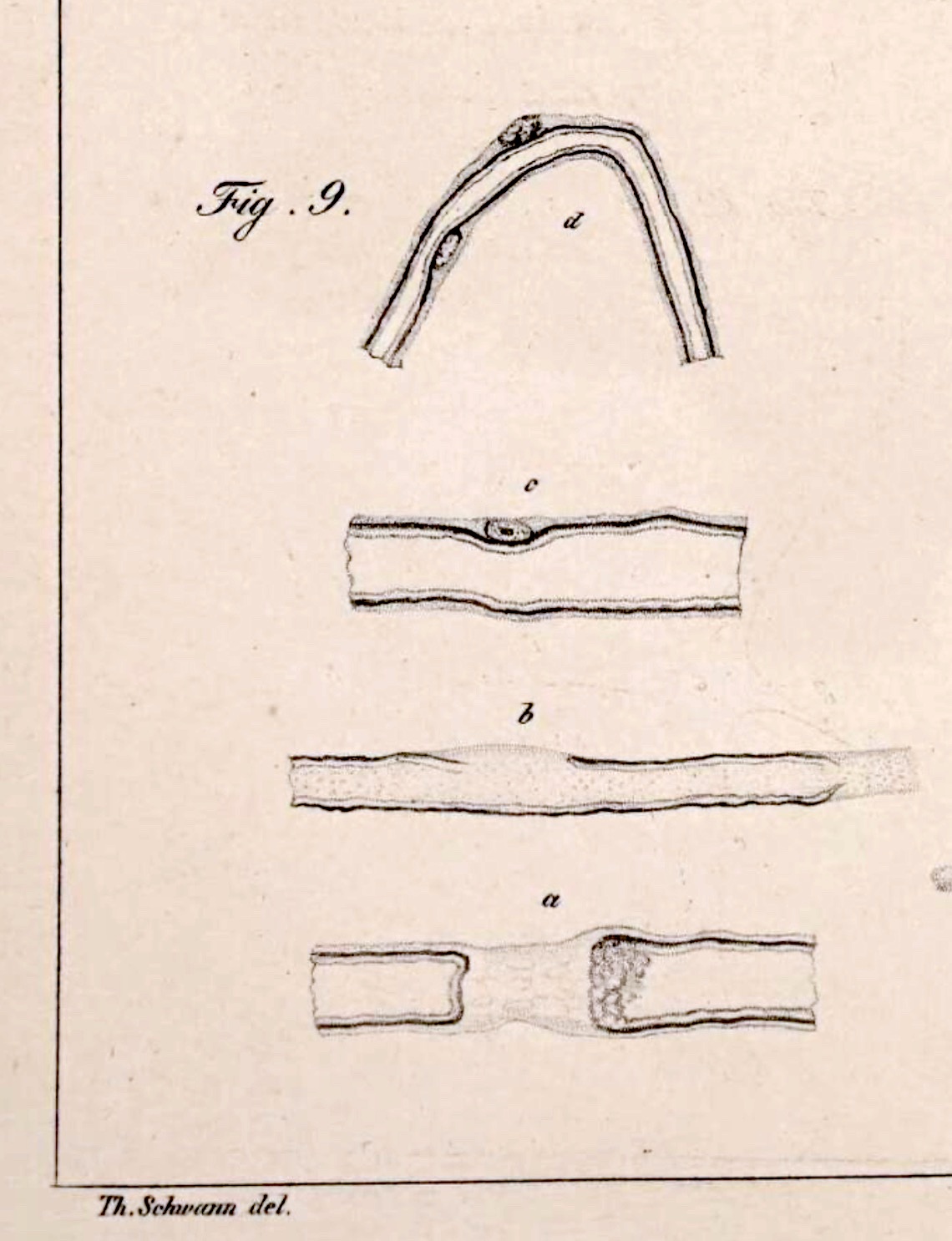 |
| Figure 9 in plate IV, from Schwann's Mikroskopische Untersuchungen |
Schwann recognized that his eponymous Schwann cells (image at right) were intimately associated
with nerve fibers (Ranvier added further clarification), but it awaited the advent of electron microscopy to reveal
that myelin was actually composed of cell membrane of the Schwann cells themselves, wrapped around and
around myelinated axons.
Development of Cell Theory
We owe the word cell, as a name for ubiquitous biological structures,
to the Englishman Robert Hooke. But what Hooke had described in 1665 were
merely small empty chambers (hence "cells") that he had observed in a thin slice of dry cork.
So accustomed have modern biologists become to calling a unit of biological organization a "cell," we
seldom notice that this word is an outrageous misnomer whose principal meaning (including as used
by Robert Hooke) remains that of "small empty chamber."
Capsule histories as commonly found in biology textbooks typically credit Schwann, along with
botanist Mathias
Jakob Schleiden (1804-1881), as founders of cell theory. However, the profound realization that
the bodies of all plants and animals are comprised of cells and cell products had begun to emerge well
before Schwann's landmark 1839 publication [Mikroskopische Untersuchungen über die Uebereinstimmung
in der Struktur und dem Wachsthum der Thiere und Pflanzen / "Microscopic Studies on the Correspondence in the
Structure and Growth of Animals and Plants"], through the efforts of
many microscopists. (Bichat, although he had founded the discipline
of histology in 1802, had not used a microscope and applied the word "cellular" ["cellulaire"] not in reference to
"cells" as eventually understood but as a label for loose areolar connective
tissue, most of which is extracellular.)
Notable pioneers in the understanding of cells include
John Goodsir (1814-1867), who... and botanist Robert Brown (1773-1858) who in 1831 described nuclei in plant cells and provided the label "nucleus" that we continue to use.
Goodsir essay
Capsule histories in biology textbooks commonly credit both Mathias
Jakob Schleiden (1804-1881) and Theodor Schwann as the originators of Cell Theory. Schleiden, a botanist,
had the easier half of this generalization, since every plant cell is encased in a durable extracellular wall
(i.e., those "cells" first reported by Robert Hooke), with very little other extracellular material to distract the observer.
Animal cells, in contrast, not only come in a confusing variety of sizes and shapes but are also associated with considerable amounts of various
extracellular materials such as collagen and ground substance. It is Schwann who receives recognition for merging his own observations of animal cells with Schleiden's
observations of plant cells, to arrive at the broad generalization that we now know as Cell Theory. Schwann's 1839 book, Mikroskopische Untersuchungen über die Uebereinstimmung in der Struktur und
dem Wachsthum der Thiere und Pflanzen ["Microscopic Studies on the Correspondence in the
Structure and Growth of Animals and Plants"] is one of the foundational works of modern biology.
However, neither Schleiden nor Schwann had properly appreciated that cells originate by division of pre-existing cells. others made significant contributions along the way, including John Goodsir and Rudolf Virchow.
The unifying observation
for Cell Theory was the presence of a "nucleus" (so named by botanist Robert Brown in 1831) within each cell of both plants and animals.
"In the course of his [Schwann's] verifications of the cell theory, in which he traversed the whole field of histology, he proved the
cellular origin and development of the most highly differentiated tissues... His generalization became the foundation of modern histology,
and in the hands of Rudolph Virchow (whose cellular pathology was an inevitable deduction from Schwann) afforded the
means of placing modern pathology on a truly scientific basis." [Quoted from The Encyclopedia Britannica's eleventh edition
(1911; vol. 24, p. 388). This classic 1911 edition is accessible through several online sources, including
here, at Wikisource.]
Additional information:
On Theodor Schwann:
[More, from Wikipedia]
[More,
from Annals of Clinical & Laboratory Science]
[More,
from Britannica]
On Cell Theory:
[More, from Wikipedia]
[More, from Britannica]
The full text of Mikroskopische Untersuchungen, with plates of Schwann's drawings,
is available at The Wellcome Collection.
A fascinating account of observations over several decades that led up to and beyond Schwann's understanding of Schwann cells can be found in:
Axel Karenberg, Schwann Cells (Ch. 7, pp. 44-50, in Neurological Eponyms, P. J. Koehler et al., eds., Oxford
University Press, 2000), available through Google Books here
(enter "Schwann" in the "Search inside" window).
A B C D
E F G H
I J K L
M N O P
Q R S T
U V W X
Y Z
TOP OF PAGE / ALPHABETICAL INDEX / CHRONOLOGICAL INDEX
Enrico Sertoli (1842-1910)
Italian histologist and physiologist, commemorated in Sertoli cells (support cells of
the testis).
For much of his career, Sertoli was professor of anatomy and physiology at the Advanced Royal School of Veterinary
Medicine in Milan, where he founded the Laboratory of Experimental Physiology.
The eponymous cells were described in Dell'esistenza di particolari cellule ramificate nei canalicoli seminiferi del testicolo umano
(About the existence of special branched cells in the seminiferous tubules of the human testis), Morgagni (1865), vol. 7, pp. 31-33.
Sertoli pursued further investigation into testicular histology, during an era when understanding the formation of reproductive
cells was of central importance in biology. In Sulla struttura dei canalicoli seminiferi dei testicoli studiata in rapporto allo
sviluppo dei nemaspermi (The structure of seminiferous tubules and the development of sperm),
Archivio per le scienze mediche (1878), he concluded (correctly, in opposition to von Ebner) that sperm cells
derived from spermatogonia rather than from Sertoli cells, which provide support.
A nice historical study of the differing interpretations of von Ebner and Sertoli may be found in the
following two papers, which include detailed annotations of the original reports:
Jones SL, Harris K, Geyer CB. A new translation
and reader's guide to Victor von Ebner's classical description of spermatogenesis. Mol. Reprod. Dev.
2019; 86:1462-1484. https://doi.org/10.1002/mrd.232821484.
Geyer, C. B. (2018). A historical perspective
on some "new" discoveries on spermatogenesis from the laboratory of Enrico Sertoli in 1878. Biology of Reproduction,
99(3), 479-481. https://doi.org/10.1093/biolre/iox125.
Additional resources:
A B C D
E F G H
I J K L
M N O P
Q R S T
U V W X
Y Z
TOP OF PAGE / ALPHABETICAL INDEX / CHRONOLOGICAL INDEX
Alexander Skene (1837-1900)
Scottish gynecologist, commemorated in Skene's glands in the female genital-urinary tract.
Brief biography at Wikipedia.
A B C D
E F G H
I J K L
M N O P
Q R S T
U V W X
Y Z
TOP OF PAGE / ALPHABETICAL INDEX / CHRONOLOGICAL INDEX
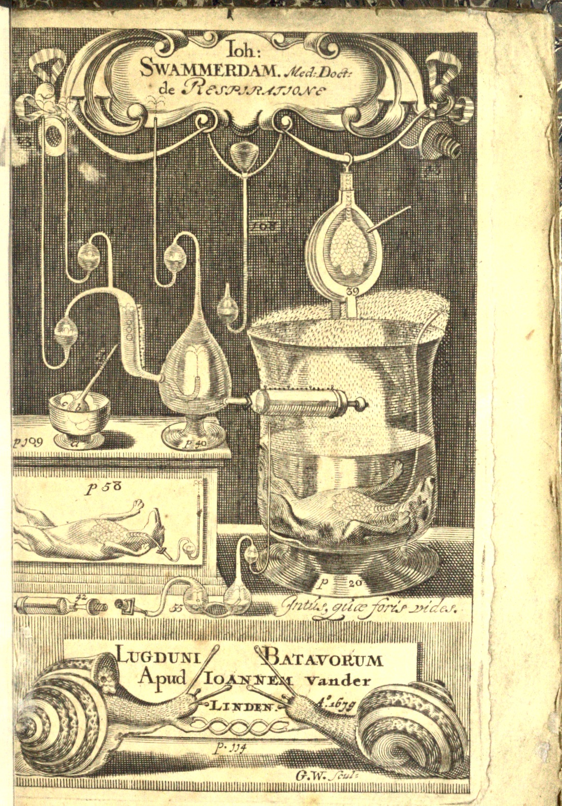 |
Frontispiece from Swammerdam's Tractatus physico-anatomico-medicus de respiratione usuque pulmonum
(A physico-anatomical-medical treatise on respiration and the use of the lungs).
[Accessed at the Wellcome Collection.]
|
Jan Swammerdam (1637-1680)
Dutch naturalist, microscopist, and physiologist, commemorated in Swammerdam valves in lymphatic
vessels.
Swammerdam trained in medicine at the University of Leiden, Netherlands. But instead of medical practice, he pursued a career of
research in anatomy, physiology, and natural history. He made his own single-lens (glass bead) microscopes and developed tools and
techniques for delicate dissections and injection of fluids, enabling visualization of lymphatic vessels (among other things). He
debunked Decarte's hydraulic theory of muscle action, by demonstrating that muscle volume does not change substantially during muscular contraction.
Swammerdam is perhaps best known for documenting the different life stages of insects (i.e., the metamorphosis of caterpillars into
moths or butterflies, grubs into beetles, maggots into flies), as well as their internal anatomy, all with finely detailed illustrations.
Additional resources:
History of lymphatic system research:
- Natalie, Bocci, & Ribatti, "Scholars and scientists in the history of the lymphatic system,"
Journal of Anatomy 231:417-429 (2017),
free access article doi: 10.1111/joa.12644.
- Loukas et al. (2011) The lymphatic system: a historical perspective. Clin Anat 24, 807-816.
A B C D
E F G H
I J K L
M N O P
Q R S T
U V W X
Y Z
TOP OF PAGE / ALPHABETICAL INDEX / CHRONOLOGICAL INDEX
Rudolf Ludwig Carl Virchow (1821-1902)
German pathologist and politician, "the father of modern pathology," commemorated in Virchow-Robin space
(perivascular extension of subarachnoid space in the brain).
Earning his doctorate soon after cells had been recognized as a fundamental unit of living
things, Virchow readily adopted the cellular perspective. It was he who popularized the dictum, "omnis cellula
e cellula": all cells come from cells. Furthermore, all diseases result from disorders in cellular function.
These premises were advocated by Virchow in the middle of the nineteenth century, soon after establishment of Cell
Theory.
"Life itself is but the expression of a sum of phenomena,
each of which follows the ordinary physical and chemical laws. ... Disease
is not something personal and special, but only a manifestation of life
under modified conditions, operating according to the same laws as apply
to the living body at all times, from the first moment until death."
(This quotation is attributed to Virchow on many webpages, but typically without crediting an original source.)
As an educator during a time when microscopes were a scarce resource, Virchow reportedly
sent his own microscope and slides from student to student on a specially-built model train.
The following text is excerpted, with ellipses and other light editing, from the 11th edition of The Encyclopedia Britannica, volume 28.
[This classic 1911 edition is accessible through several online sources, including
here, at Wikisource.]
"[Virchow] took his doctor's degree in 1843, and almost immediately received an appointment as assistant-surgeon at the Charité Hospital... In 1847 he founded with Reinhardt the Archiv für pathologische Anatomie [later known as Virchow's Archiv]... In 1848 he went as a member of a government commission to investigate an outbreak of typhus in upper Silesia. About the same time, having shown too open sympathy with the revolutionary or reforming tendencies of 1848, he was for political reasons obliged to leave Berlin... In 1856 he was recalled to Berlin..., and as director of the Pathological Institute formed a centre for research whence has flowed a constant stream of original work on the nature and processes of disease.
"Wide as were Virchow's studies, and successful as he was in all, yet the foremost place must be given to his achievements in pathological investigation. He may, in fact, be called the father of modern pathology, for his view, that every animal is constituted by a sum of vital units, each of which manifests the characteristics of life, has almost uniformly dominated the theory of disease since the middle of the 19th century, when it was enunciated...
"Virchow made many important contributions to histology and morbid anatomy and to the study of particular diseases. The [tissue-based] classification into epithelial organs, connective tissues, and the more specialized muscle and nerve, was largely due to him... [emphasis added; c.f., Bichat]
"Another science which Virchow cultivated with conspicuous success was anthropology, which he did much to put on a sound critical basis. At the meeting of the Naturforscherversammlung at Innsbruck in 1869, he was one of the founders of the German Anthropological Society... His archaeological work included the investigation of lake dwellings and other prehistoric structures; he went with Schliemann to Troy in 1879, ... and in 1888 he accompanied Schliemann to Egypt...
"As a politician Virchow had an active career. ...[T]he expression Kulturkampf ["culture struggle"] had, it is believed, its origin in one of his electoral manifestoes... As a member of [Berlin's] municipal council [Virchow] was largely responsible for the transformation which came over the city in the last thirty years of the 19th century. That it has become one of the healthiest cities in the world from being one of the unhealthiest is attributable in great measure to his insistence on the necessity of sanitary reform, and it was his unceasing efforts that secured for its inhabitants the drainage system, the sewage farms and the good water-supply, the benefits of which are reflected in the decreased death-rate they now enjoy...
"[Virchow's] eightieth birthday was celebrated in Berlin amid a brilliant gathering of men of science, part of the ceremonies taking place in the new Pathological Museum, ... which owes its existence mainly to his energy and powers of organization. On that occasion all Europe united to do him honour..."
Virchow's efforts to advance a scientific approach to medicine are eloquently described in a hagiographic essay published during his lifetime,
in The Popular Science Monthly, Oct. 1882, pp. 836-842. This essay should be available at Google Books, here.
Another encomium reviewing Virchow's career was published in 1921
(C.V. Weller, "Rudolf Virchow--Pathologist,"The Scientific Monthly, Vol. 13, pp. 33-39).
The Virchow entry at Wikipedia is quite extensive and includes an account
of Virchow's opposition to Darwinism. This entry also identifies as apocryphal the oft-told tale of a sausage duel between Virchov and German Chancellor Otto von Bismark.
A nineteenth-century controversy regarding John Goodsir's contribution to cell theory, including accusations that Virchow had inadequately credited Goodsir, is ably discussed here, by K. Donaldson1 & C. Henry in the Journal of the Royal College of Physicians of Edinburgh (2020) 50: 188-195. (The authors exonerate Virchow.)
The interested reader is additionally directed to the
text of a dramatic monologue which ably presents Virchow's life and times.
This monologue, presented by Ed "The Pathguy" Friedlander, M.D., to a meeting
of the Group for Research in Pathology Education, includes numerous direct quotations from the historical Dr. Virchow, as well as a list of
print resources.
A B C D
E F G H
I J K L
M N O P
Q R S T
U V W X
Y Z
TOP OF PAGE / ALPHABETICAL INDEX / CHRONOLOGICAL INDEX
Anton Gilbert Victor von Ebner (1842-1925)
Austrian histologist, commemorated in von Ebner's glands (salivary glands in tongue)
and von Ebner's lines (growth lines in teeth).
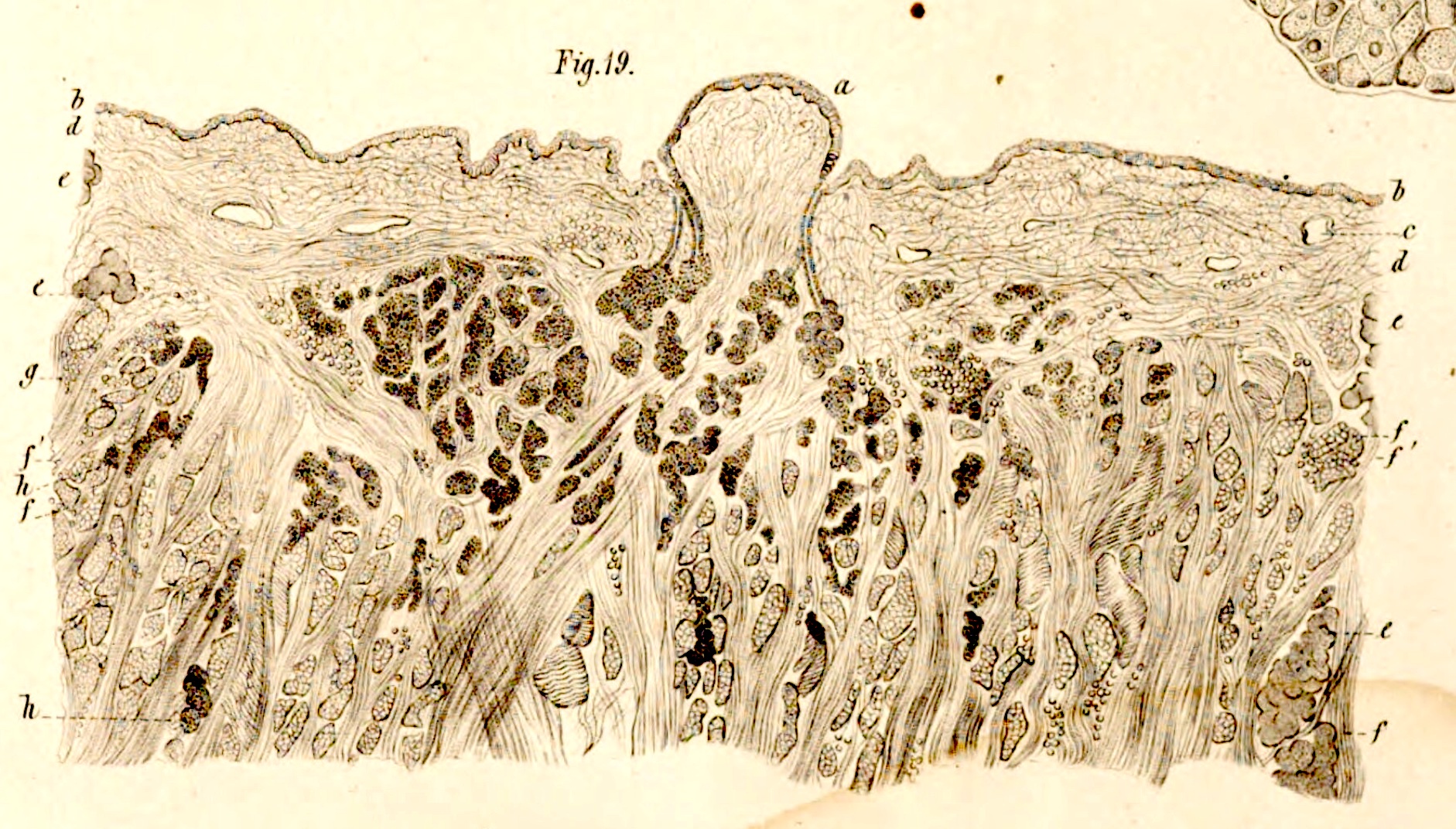 |
| Tissues of tongue from Die acinösen
Drüsen der Zunge, 1873, showing von Ebner's glands beneath a
circumvallate papilla.
|
The eponymous salivary glands
of the tongue are described in Die acinösen
Drüsen der Zunge und ihre Beziehungen zu den Geschmacksorganen:
eine anatomische Untersuchung [The acinar glands of the tongue and their relations to the organs of taste: an anatomical study],
University of Graz, 1873.
Von Ebner received his medical degree from the University of Vienna in 1866. He became professor of histology at the
University of Graz, later at the University of Vienna. He edited the third volume of the sixth edition (1899) of
Kölliker's Handbuch der Gewebelehre des Menschen (Manual of human histology).
In a paper which he submitted in 1871 in consideration of promotion and tenure, von Ebner made substantial contribution to
understanding spermiogenesis. This paper, Untersuchungen über den Bau der
Samencanälchen und die Entwicklung der Spermatozoiden [Studies of the Structure of the Seminiferous Tubules and the
Development of Sperm], appeared at a time when understanding the formation of reproductive
cells was of central importance in biology. Although von Ebner wound up on the wrong side of history by interpreting Sertoli
cells as the source of spermatozoa, his work was impressively detailed and accurate given technical limitations of his era.
A nice historical study of the differing interpretations of von Ebner and Sertoli may be found in the
following two papers, which include detailed annotations of the original reports:
Jones SL, Harris K, Geyer CB. A new translation
and reader's guide to Victor von Ebner's classical description of spermatogenesis. Mol Reprod Dev.
2019; 86:1462-1484. https://doi.org/10.1002/mrd.232821484.
Geyer, C. B. (2018). A historical perspective
on some "new" discoveries on spermatogenesis from the laboratory of Enrico Sertoli in 1878. Biology of Reproduction, 99(3), 479-481.
https://doi.org/10.1093/biolre/iox125.
Wikipedia offers a very brief biography, with a list of publications.
A B C D
E F G H
I J K L
M N O P
Q R S T
U V W X
Y Z
TOP OF PAGE / ALPHABETICAL INDEX / CHRONOLOGICAL INDEX
Heinrich Wilhelm Gottfried von Waldeyer-Harz (1836-1921)
German anatomist, commemorated in Waldeyer's ring of circumpharyngeal lymphoid tissue. Waldeyer is especially noted for introducing the words chromosome and neuron into the vocabulary of biology. By summarizing Cajal's work, Waldeyer helped to establish the Neuron Doctrine
As a student at the University of Göttingen (1857), Waldeyer was inspired by lectures of Jakob Henle to direct his studies into anatomy. After obtaining his doctorate in Berlin in 1861, he worked at the University of Königsberg and the University of Breslau before settling in Strasbourg (where Germans were being installed to replace French professors after the conquest of Alsace by Prussia in 1871). He remained in Strasbourg for eleven years before moving again to the Charité hospital associated with the University of Berlin, where he remained for 33 years until his retirement in 1916.
Waldeyer was famed not only for the breadth of his own anatomical research but for his interpretations of results accumulated by others, especially in areas of neuroanatomy and cytology. He was also known for the excellence of his teaching, which attracted students from across the world (e.g., Lanterman). But he was infamously opposed medical education for women.
Near the end of his life (1921), Waldeyer wrote a lengthy autobiography, Lebenserinnerungen, or "life memories." (The link should take you to an incomplete version at GoogleBooks.)
Among Waldeyer's principal monographs are:
Über Karyokinese und ihre Beziehungen zu den Befruchtungsvorgängen ("About Karyokinesis and its relationships with the fertilization processes"), Archiv für mikroskopische Anatomie und Entwicklungsmechanik, 32: 1-122 (1888).
Eierstock und Ei-Ein Beitrag zur Anatomie und Entwicklungsgeschichte der Sexualorgane ("Ovary and Egg - A contribution to the anatomy and developmental history of the sexual organs"), Leipzig, W. Engelmann (1870).
Das Becken-Topografisch-anatomisch, mit besonderer Berücksichtigung der Chirurgie und Gynäkologie ("The pelvis-topographic-anatomical, with special attention to surgery and gynaecology"), a continuation of Johann Georg Joessel's (1838-1892) Lehrbuch der topgrafisch-chirurgischen Anatomie. Volume II, Bonn (1899).
Über einige neuere Forschungen im Gebiete der Anatomie des Zentralnervensystems ("On some recent research in the field of anatomy of the central nervous system"), Dtsch. Med. Wochenschr. 17:1213-1218 (1891).
Whonamedit.com includes a more extensive listing of Waldeyer's publications.
For essays about Waldeyer, see:
Wilhelm Waldeyer--An Important Scientific Researcher of the 19th Century in the Context of His Memoirs and Major Monographies, H. Scheuerlein, C. Pape-Köhler, & F. Köckerling, Front. Surg. 8, DOI 10.3389/fsurg.2021.752709 (2021). This article summarizes Waldeyer's Lebenserinnerungen as well as some of his principal anatomical works.
Wilhelm von Waldeyer-Hartz -- A Great Forefather: His Contributions to Anatomy with Particular Attention to "His" Fascia, H. Scheuerlein, F. Henschke, F. Köckerling, Front Surg. 4: 74 DOI: 10.3389/fsurg.2017.00074 (2017).
Centennial of Wilhelm Waldeyer's introduction of the term "chromosome" in 1888, T. Cremer & C. Cremer, Cytogenet. Cell Genet. 48:66-67 (1988).
Wikipedia offers a bit of additional information.
A B C D
E F G H
I J K L
M N O P
Q R S T
U V W X
Y Z
TOP OF PAGE / ALPHABETICAL INDEX / CHRONOLOGICAL INDEX
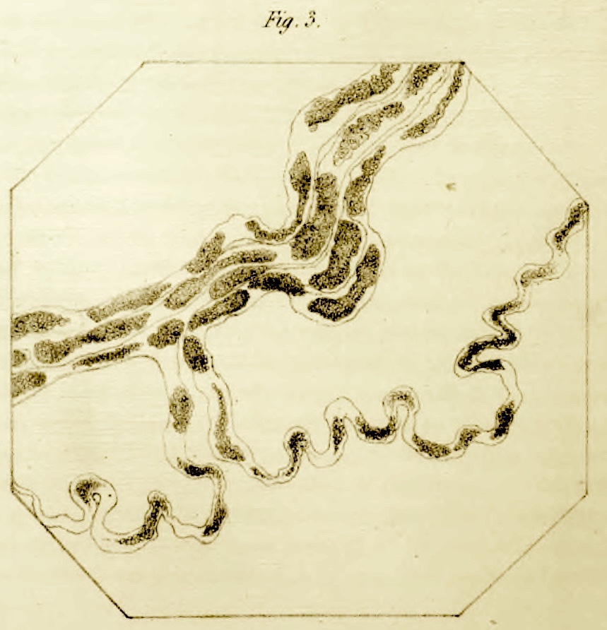 |
Image from Phil. Trans. Roy.
Soc. London (1850):
"Fig. 3. Disorganized muscular nerve, from the inferior surface of the tongue, five days after
section." |
Augustus Volney Waller (1816-1870)
English physician, commemorated in Wallerian (or anterograde)
degeneration, whereby nerve fibers begin to fall apart after separation from their cell bodies. This response proved invaluable over
subsequent decades for mapping neural pathways, in the discipline founded by Waller that became known as experimental neurology.
After earning his medical degree in Paris, Waller acquired his British medical license in 1840 and established a general practice in Kensington. In 1851 he
left England to pursue research in Bonn, subsequently continuing his research in Paris.
The following excerpts are from the
Dictionary of National Biography (1885-1900), at Wikisource:
"Waller was endowed with a remarkable aptitude for original investigation. Quick to perceive new and promising lines of research,
and happy in devising processes for following them out, he possessed consummate skill ... in experimental work. His discoveries
in connection with the nervous system constitute his most conspicuous claim to distinction, and the fields he first traversed have proved
fruitful beyond imagination, for they have led directly to nearly all that we know experimentally of the functions of the nervous system.
"His ... name will long be associated with the degeneration method of studying the paths of nerve impulses, for he invented it.
He did not confine himself to a consideration of the nervous system, however, for he practically rediscovered the power which the white
blood corpuscles possess of escaping from the smallest blood-vessels..."
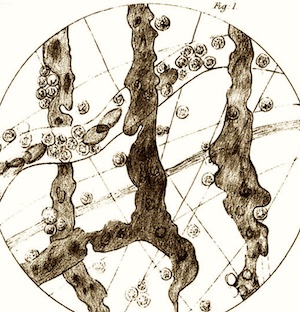 |
Image from Philosophical Magazine (1846):
"Fig.1... vessels of the inferior surface of the tongue as they appear after the escape of the corpuscles... A portion of a vessel with an
internal current is likewise seen with discs, and internal and external corpuscles..." |
Waller's account of the extravasation of white blood cells (illustration at right) was published in 1846:
"Microscopic Observations on the Perforation
of the Capillaries by the Corpuscles of the Blood and on the origin of mucus and pus globules,
Philosophical Magazine vol. 29, pp. 397-405. This report came just a few years after establishment of Cell Theory by
Schwann, well before the nature of capillaries had become thoroughly understood.
(For a more thorough account of historical understanding of capillaries, see
"The history of the capillary wall:
doctors, discoveries, and debates," by C. Hwa and W.C. Aird, in
Am J Physiol Heart Circ Physiol 293: H2667-H2679 (2007),
doi:10.1152/ajpheart.00704; also see
"
Completing the puzzle of blood circulation: the discovery of capillaries," from ResearchGate.)
Waller's description of the eponymous process of axonal degeneration, which acknowledged relevant prior observations by others, was read
into the Philosophical Transactions of the Royal Society of London (vol. 140:, pp. 423-429) in 1850, before he moved abroad in search of
improved research opportunities. The illustration here (above right) was taken from this report,
"Experiments on the section of the glossopharyngeal and
hypoglossal nerves of the frog, and observations of the alterations produced thereby in the structure of their primitive fibres."
Additional notes
The entry for Waller at Whonamedit.com includes an incomplete but
extensive bibliography.
According to both Wikipedia and
Whonamedit.com, Waller's son, Augustus Desiré Waller,
developed the first practical apparatus, using surface electrodes, for electrocardiography. (The Wikipedia entry for Waller himself largely reproduces the old entry in the Dictionary
of National Biography (1885-1900), cited above.)
A B C D
E F G H
I J K L
M N O P
Q R S T
U V W X
Y Z
TOP OF PAGE / ALPHABETICAL INDEX / CHRONOLOGICAL INDEX
Karl Weigert (1845-1904)
German pathologist, commemorated in Weigert stain for myelin,
also Weigert's elastic stain.
Weigert studied at the universities of Berlin, Vienna, and Breslau, served as an assistant surgeon during the Franco-Prussian War, and subsequently as
an assistant to Heinrich Waldeyer.
He is reported as the first to stain bacteria and was able to demonstrate the presence of bacteria in histological sections.
Weigert was cousin to Nobel laureate Paul Ehrlich, of "magic bullet" fame,
and teamed up with Ehrlich and Ludwig Edinger to establish the Senckenbergisches Pathologish-Anatomisches Institut in Frankfurt am Main as a top-notch
research institute.
The biography with bibliography at whonamedit.com tells us that
"Weigert's most notable personal characteristic was his excessive modesty."
Wikipedia and
the Jewish Encyclopedia
also offer brief biographies.
A B C D
E F G H
I J K L
M N O P
Q R S T
U V W X
Y Z
TOP OF PAGE / ALPHABETICAL INDEX / CHRONOLOGICAL INDEX
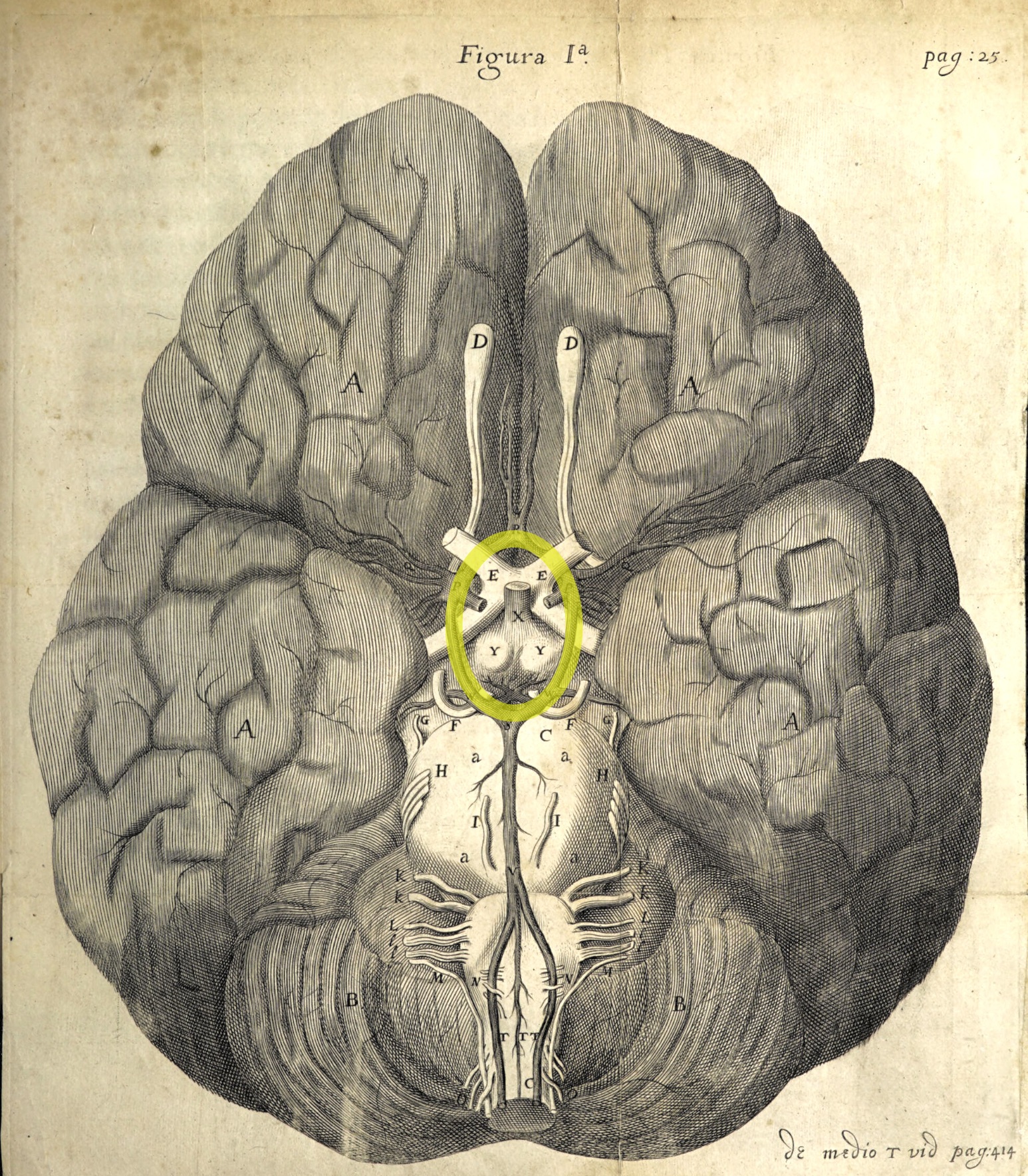 |
Illustration by Christopher Wren, in Cerebri anatome. Most features should be readily recognizable
by modern students of neuroanatomy; blood vessels comprising the circle of Willis are highlighted in yellow.
[Accessed at the Wellcome Collection.]
|
Thomas Willis ((1621-1675)
English physician, alchemist, and anatomist. Although Willis has no histological eponyms, his foundational work in neurology
eventually led to some of the most exciting developments in histology. He himself coined the word "neurologie" to
identify his new field of study, and he is commemorated in the circle of Willis, a ring of
interconnected arteries at the base of the cerebrum.
Willis's reputation as a neuroanatomist was secured by Cerebri
anatome: cui accessit nervorum descriptio et usus [Anatomy of the brain, including description and use of nerves].
This great work, published in 1664, was illustrated with drawings by Christopher Wren, who went on to become the renowned
architect of St. Paul's Cathedral in London.
In addition to Wren, others among Willis's notable contemporaries included William Harvey,
who first demonstrated the circulation of blood. Both Willis and Harvey practiced medicine and conducted research in Oxford,
even while the city was besieged by Cromwell's forces during the English Civil War (ca. 1646). Both were Royalists, with Willis
himself serving as a soldier in King Charles's army. Willis eventually joined other famous colleagues as fellows in the
newly-formed Royal Society.
Willis produced his major work at a time when Rene Decartes (1596-1650) had proposed that brain function was pneumatic, with cerebral ventricles pumping air
to inflate muscles. Contemporary British philosopher
Henry More (1614-1687) regarded brain tissue itself as no more capable of thought than "a bowl of curds." For his own studies, Willis developed improved
approaches for studying neuroanatomy, including pickling brains in spirits of wine (distilled alcohol) to render their texture less like custard and more like
boiled eggwhite. The anatomy of peripheral nerves suggested to Willis that brain function involved "vital spirits" whose energetic corpuscles
could enter the brain through sensory nerves, interact within the substance of the brain, and then travel throughout the body along motor nerves where
their energy could prompt muscle action.
In contrast to William Harvey's theory of blood circulation which was based on abundant experimental evidence (and confirmed by
Marcello Malpighi a few years after his death), Willis's ideas of brain function lacked any substantial experimental
support during his lifetime; his materialistic views would languish for centuries before the sciences of histology and neurophysiology could
together confirm the reality of complex axonal pathways carrying energetic impulses throughout the brain and body at large. Nevertheless,
even today when neuroscientists can study the neural correlates of perception, thought, and emotion, the fundamental nature of self-aware conscious
experience (i.e., "soul;" Willis's "rational spirit") remains nearly as mysterious as it was for Willis and Decartes.
Although Willis's success as a physician was not notably better than that of other doctors of his time (medical practice in the seventeenth century
had little basis in evidence, and was often harmful), he was nevertheless highly regarded.
Many of the human bodies he used for autopsy were acquired from the families of his patients, to better understand the causes of
their deaths.
Willis's fascinating life and times are much more extensively described in Carl Zimmer's captivating 2004 book,
Soul Made Flesh. (Inclusion of Willis in this Histology Eponyms
page was largely inspired by Zimmer's book.)
Additional resources:
A B C D
E F G H
I J K L
M N O P
Q R S T
U V W X
Y Z
TOP OF PAGE / ALPHABETICAL INDEX / CHRONOLOGICAL INDEX
James Homer Wright (1869-1900)
American pathologist, commemorated in Wright's stain for blood smears.
Wright identified megakaryocytes as the source for blood platelets.
Brief biography at Wikipedia.
More extensive biography in the American
Journal of Surgical Pathology, (2002) 26:88-96; doi: 10.1097/00000478-200201000-00011.
A B C D
E F G H
I J K L
M N O P
Q R S T
U V W X
Y Z
TOP OF PAGE / ALPHABETICAL INDEX / CHRONOLOGICAL INDEX
Eduard Zeis (1807-1868)
German surgeon and ophthalmologist, commemorated in glands of Zeis, sebaceous glands
associated with eyelashes. (Not to be confused with
Carl Zeiss, b. 1816, founder in 1846 of a company manufacturing fine microscopes.)
Eduard Zeis is noted primarily for publishing in 1838 the first textbook of plastic surgery,
Handbuch der plastischen Chirurgie. An English translation of this book
is available in print, as Zeis' Manual of Plastic Surgery, from Oxford University Press 1988.
For a brief biography, see Wikipedia.
A somewhat more detailed biography, listing additional publications, is available at the German language
Wikisource. (This may be readily translated
by copy-and-pasting into Google Translate.)
A B C D
E F G H
I J K L
M N O P
Q R S T
U V W X
Y Z
TOP OF PAGE / ALPHABETICAL INDEX / CHRONOLOGICAL INDEX
Johann Gottfried Zinn (1727-1759)
German anatomist and botanist, commemorated in the zonule of Zinn, the circumferential band of suspensory fibers
which anchor the lens of the eye. This is the same Johann Zinn who, as director of the botanical garden at the University of Göttingen, was recognized by Carl von Linne (Linnaeus) in the name of the flower genus Zinnia.
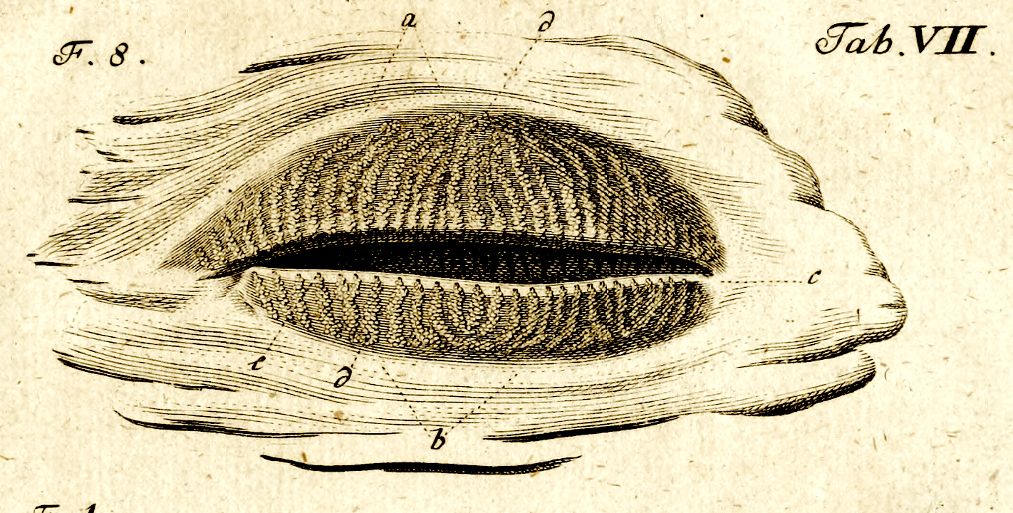 | |
Image of Meibomian glands on the inside of the eyelid
(from Zinn 1780, accessed at the Wellcome
Collection).
For a prior view of Meibom's glands, see Meibom, 1666.
|
Zinn is noted for publishing the first comprehensive atlas of eye anatomy, Descriptio Anatomica Oculi Humani Iconibus
Illustrata. Digital facsimiles,
with illustrations of Zinn's delicate dissections, may be found at the following sites:
- The Wellcome Collection displays an 1780 edition.
- The Hatha Trust offers the 1755 edition, published shortly
before Zinn's untimely death in 1759, at age 32.
An account of efforts by early anatomists to understand the suspension of the lens may be found
here [S. Bassnett (2021) Zinn's zonule.
Progress in retinal and eye research vol. 82: 100902.
doi:10.1016/j.preteyeres.2020.100902]. This article includes a discussion of the widespread but etymologically questionable
misuse of the word "zonule" as a name for individual suspensory fibers rather than for the full band of fibers which suspends the lens.
For a brief biography with bibiliography, see Whonamedit.com.
The bio at Wikipedia is extremely brief, but it does
include a photo of a Zinnia.
A B C D
E F G H
I J K L
M N O P
Q R S T
U V W X
Y Z
TOP OF PAGE / ALPHABETICAL INDEX / CHRONOLOGICAL INDEX
Chronological index by birth-year
(Alphabetical index)
Boldface highlights entries who are especially noteworthy
in the history of histology.
Italics indicates entries who are not eponyms but are included for their relevance to histology.
TOP OF PAGE / ALPHABETICAL INDEX
Copyright info: We believe that images used at this website are in the public domain. If you believe that you own copyright to any image used here, please contact us at dgking@siu.edu and we shall remove the image or add an acknowledgement.
Comments and questions: dgking@siu.edu
SIUC / School
of Medicine / Anatomy / David
King
https://histology.siu.edu/eponyms.htm
Last updated: 20 January 2026 / dgk
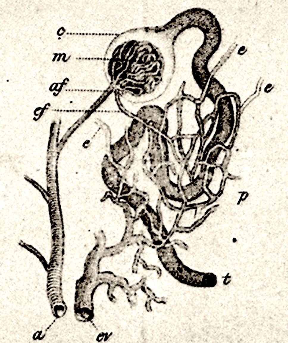
 Leopold Auerbach (1828-1897)
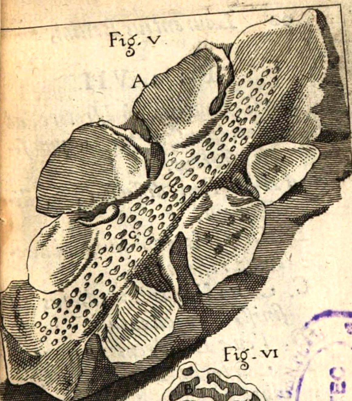

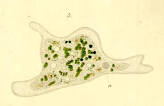
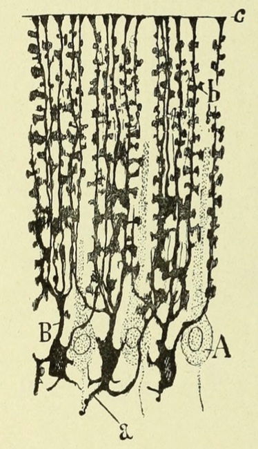
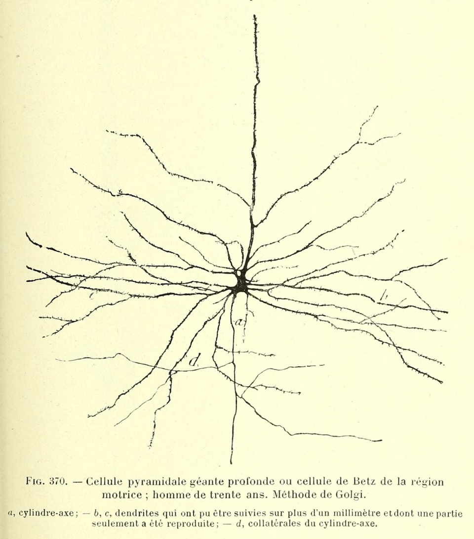
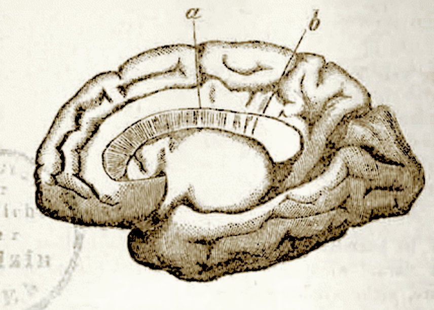
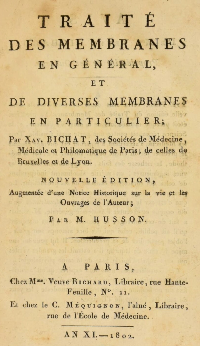
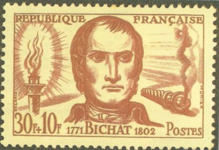
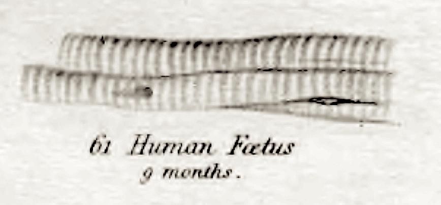
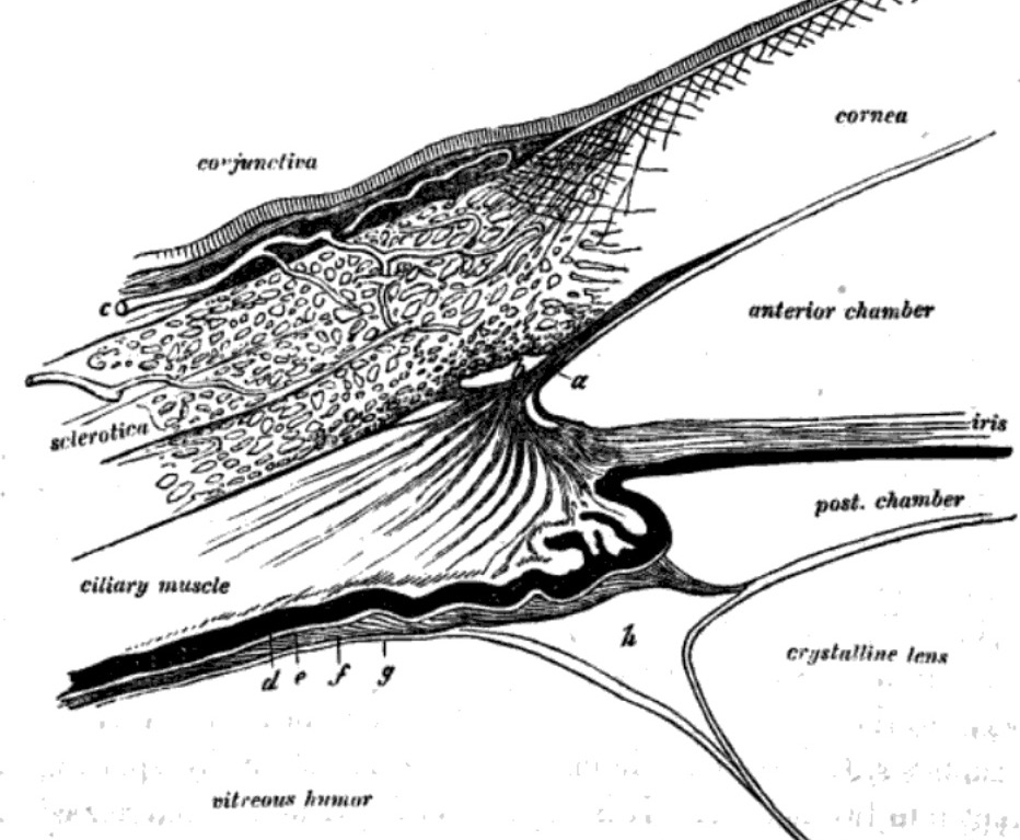
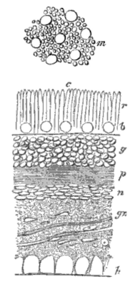
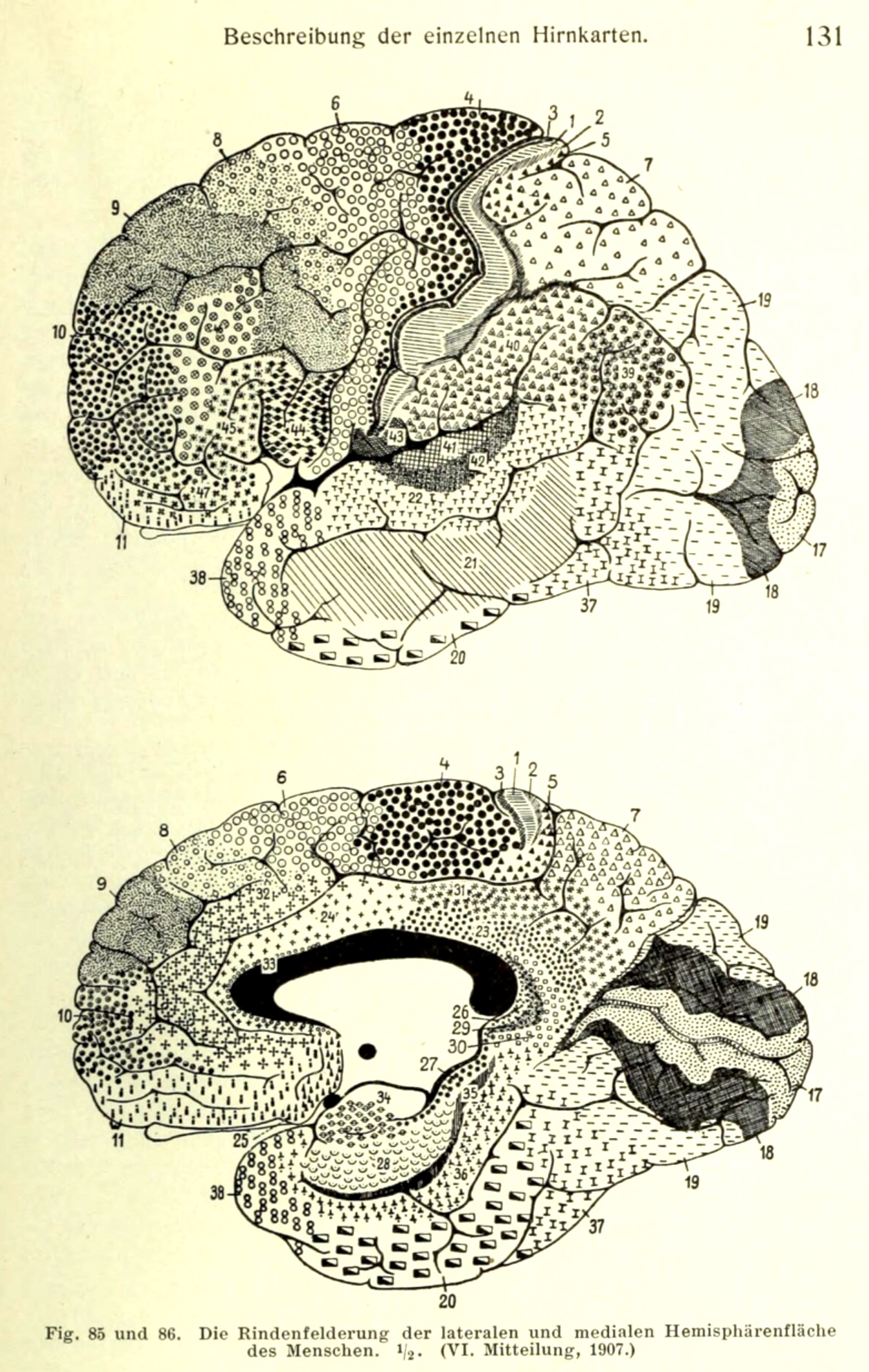
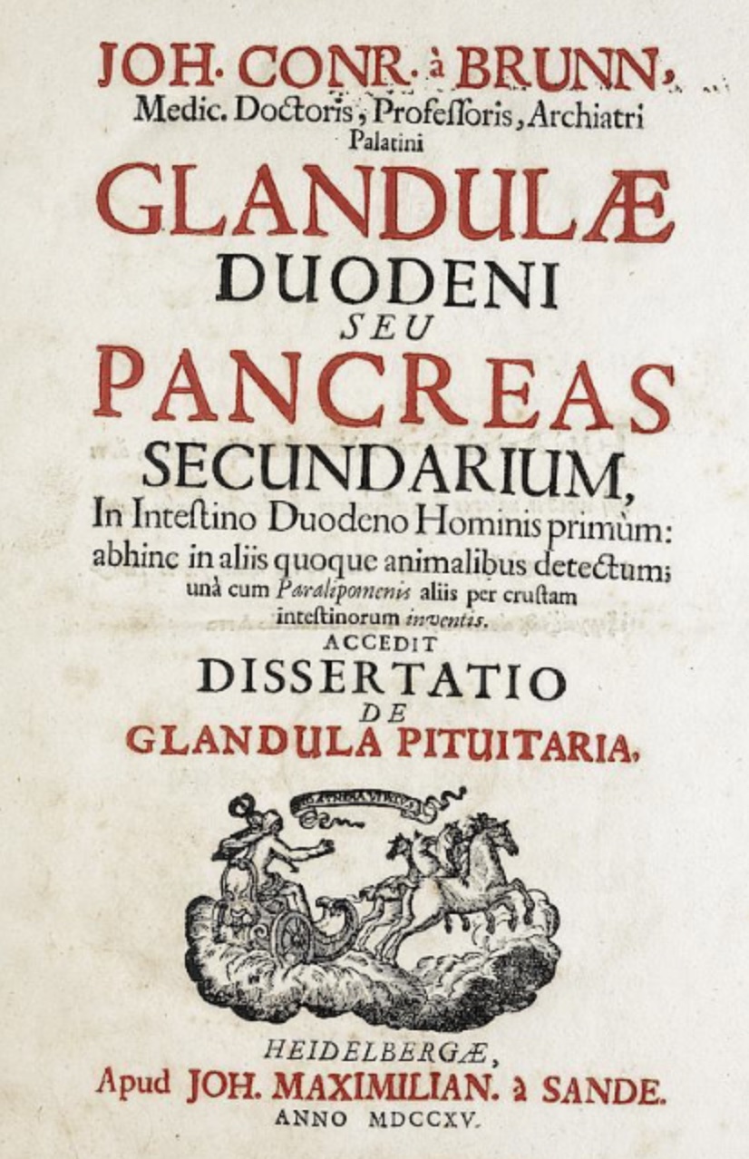
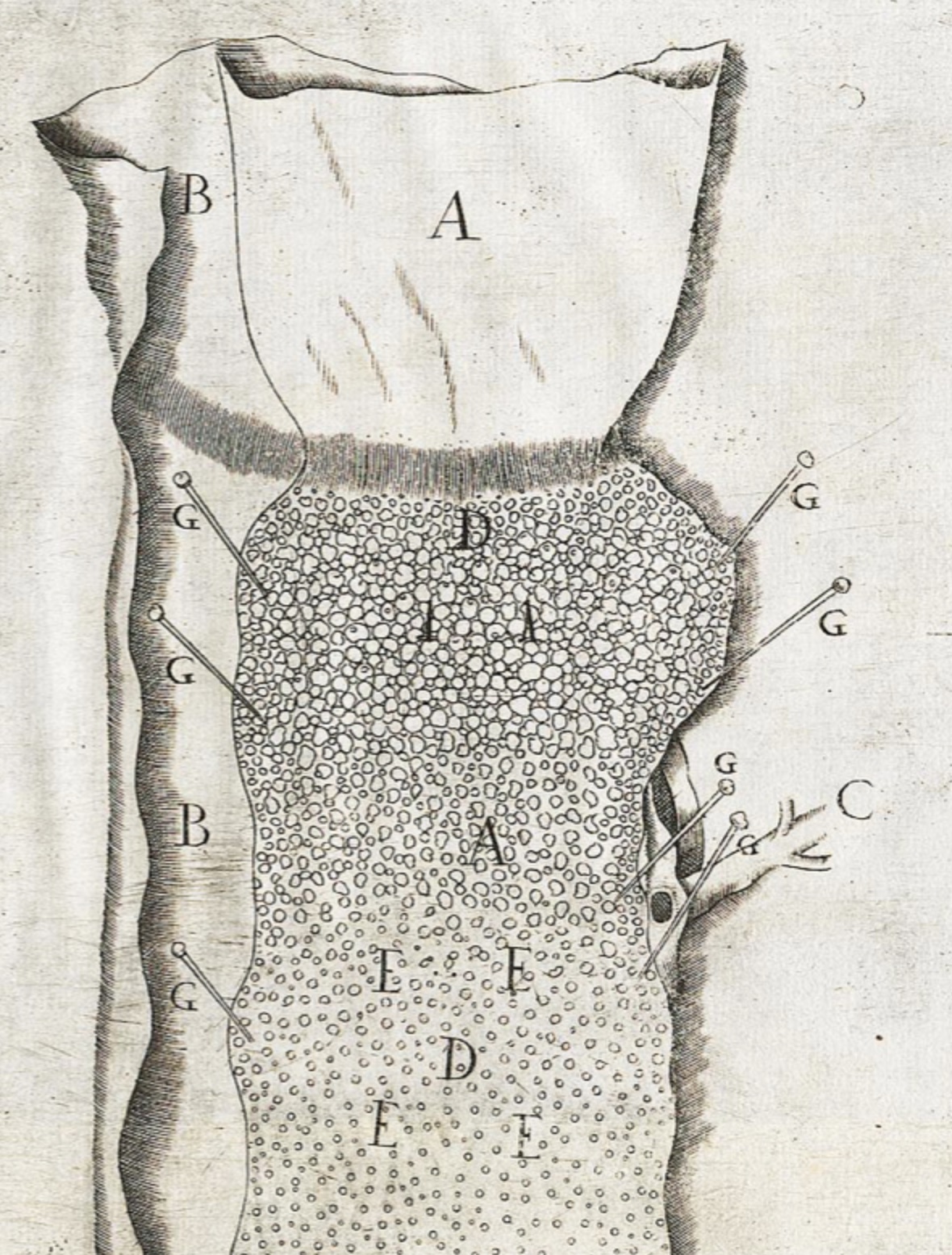
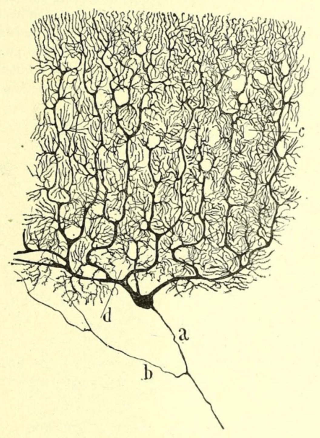


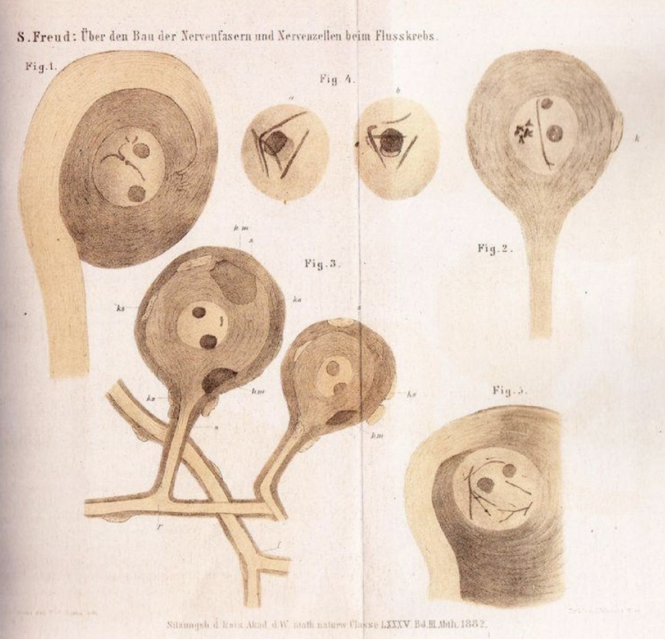


 This letter, Exercitatio Anatomica de Motu Cordis et
Sanguinis in Animalibus [


 Theodor Kerckring (ca. 1638-1693)


 August Franz Joseph Karl Mayer (1787-1865)
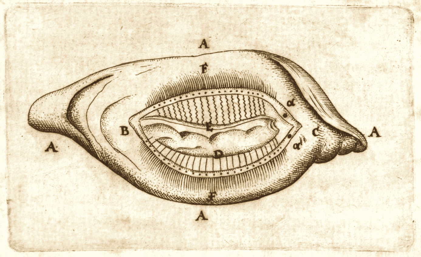
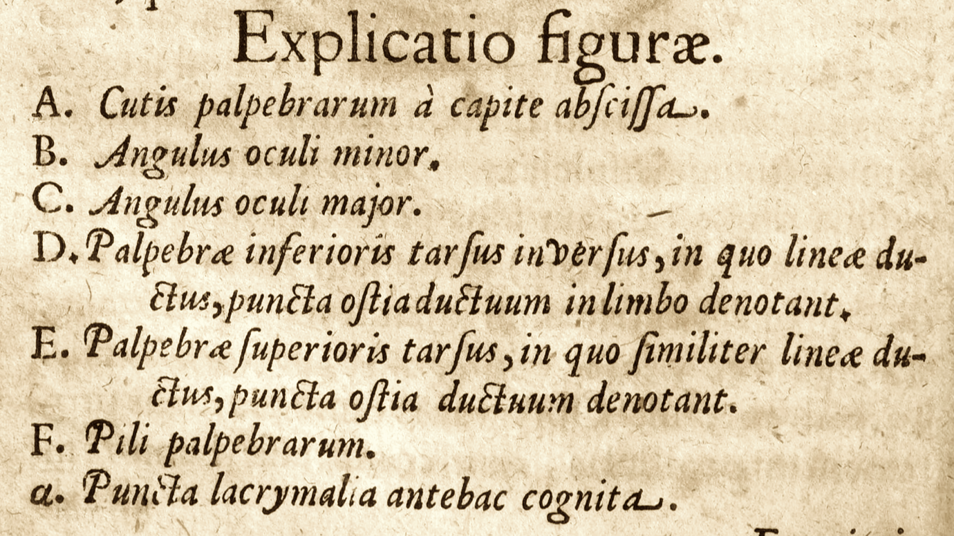


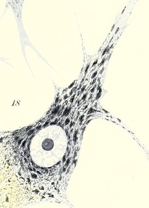


 Josef Paneth (1857-1890)
 Jan (or Johann) Evangelista Purkinje (1787-1869)
 Louis Antoine Ranvier (1835-1922)


