
Renal Corpuscle, in renal cortex
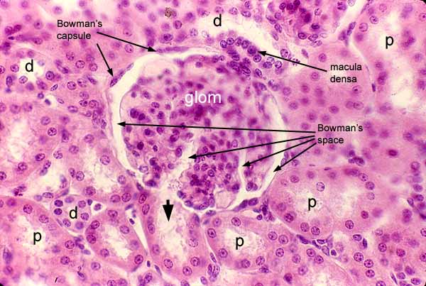
The cortex of the kidney is distinguished by characteristic renal corpuscles, each of which consists of an outer envelope of simple squamous epithelium (Bowman's capsule) surrounding a fluid-filled space (Bowman's space) within which is suspended a glomerulus (glom).
The urinary pole of this corpuscle is marked by the beginning of the proximal tubule (arrowhead), which receives filtrate from Bowman's space.
The macula densa marks the vascular pole on the other side of this corpuscle, displaying its characteristic appearance of several distal tubule nuclei crowded densely together.
Although the glomerulus in this image clearly contains many cells with varied appearances, their individual identities as endothelial cells, podocytes, or mesangial cells are difficult to determine reliably on relatively thick-sectioned specimens such as this.
The bulk of the cortex consists of convoluted tubules. Cells comprising proximal tubules (p) stain more intensely eosinophilic than those comprising distal tubules (d). The lumens of distal tubules (d) commonly appear more open and clear than those of proximal tubules (p).
Because the proximal convoluted tubule is considerably longer than the distal convoluted tubule, a typical section of the renal cortex includes many more profiles of proximal tubules than of distal tubules.
Comments and questions: dgking@siu.edu
SIUC / School
of Medicine / Anatomy / David
King
https://histology.siu.edu/crr/RN003b.htm
Last updated: 29 May 2022 / dgk