
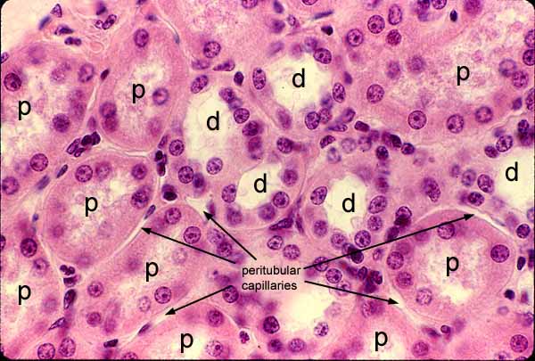
The bulk of the renal cortex consists of convoluted tubules. Cells comprising proximal tubules (p) stain more intensely eosinophilic than those comprising distal tubules (d) and have nuclei spaced somewhat farther apart. The lumens of distal tubules (d) commonly appear more open and clear than those of proximal tubules (p).
Because the proximal convoluted tubule associated with a nephron is considerably longer than the associated distal convoluted tubule, a typical section of the renal cortex includes many more profiles of proximal tubules than of distal tubules.
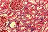
Cortical stroma, including peritubular capillaries, is generally inconspicuous. The dense, flattened nuclei in this image are those of stromal fibroblasts and capillary endothelium. Stromal collagen can be visualized with trichrome stain: Click thumbnail to right.
Click on the thumbnail at left for self-evaluation of tubule recognition.
Comments and questions: dgking@siu.edu
SIUC / School
of Medicine / Anatomy / David
King
https://histology.siu.edu/crr/RN008b.htm
Last updated: 30 May 2022 / dgk