
RENAL IMAGE INDEX / RENAL STUDY GUIDE
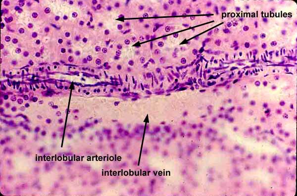
Here an interlobular arteriole lies alongside an interlobular vein, within the renal cortex.
Note the distinctive appearance of endothelial and smooth muscle nuclei in this longitudinally-sectioned arteriole. Where the lumen of the arteriole lies within the plane of section, endothelial nuclei lie along the edge of the lumen, while smooth muscle nuclei appear small and round (detail at right). Where the wall of the arteriole lies within the plane of section, smooth muscle nuclei appear much longer as their fibers wrap around the arteriole.
For a completely different view of renal cortical vasculature, click on the thumbnail at left.
Comments and questions: dgking@siu.edu
SIUC / School
of Medicine / Anatomy / David
King
https://histology.siu.edu/crr/RN043b.htm
Last updated: 22 May 2022 / dgk