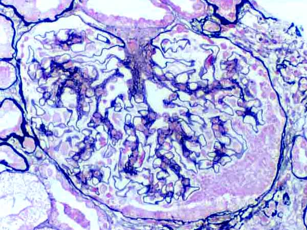
Renal biopsy, silver stain
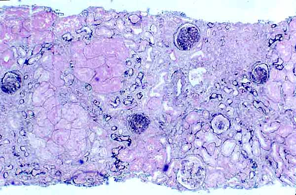
RENAL IMAGE INDEXThis low-magnification micrograph shows the full width of a needle-biopsy of renal cortex, from a patient with glomerulosclerosis. This specimen has been stained with silver to demonstrate filtration membranes and mesangial matrix. (The length of the actual specimen is several times longer than shown here.)
Mesangial occlusion of several glomeruli is fairly evident even at this low magnification, as is pathological shrinkage and degeneration of renal tubules.
Below are several higher-magnification images, illustrating variations in glomerular and tubular pathology found within this single specimen.
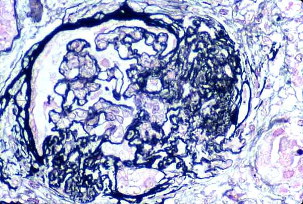
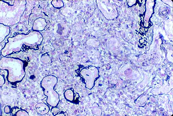
Corpuscle with patent capillaries and Bowman's space, but with significant increase in mesangial cells and matrix. Click on image for enlarged view.
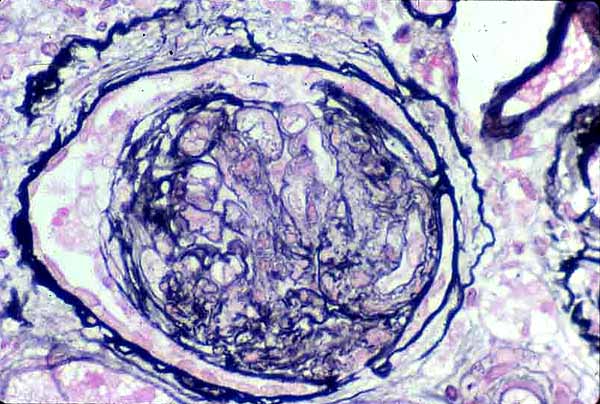
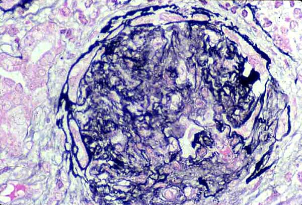
Glomerulus completely occluded, Bowman's space still partially open.
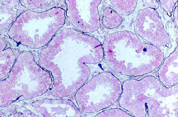
More-or-less normal renal tubules (some thickening of basement membranes). Click on image for enlarged view.
Comments and questions: dgking@siu.edu
SIUC / School
of Medicine / Anatomy / David
King
https://histology.siu.edu/crr/RN078b.htm
Last updated: 15 September 2021 / dgk