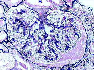
Renal corpuscle, silver stain
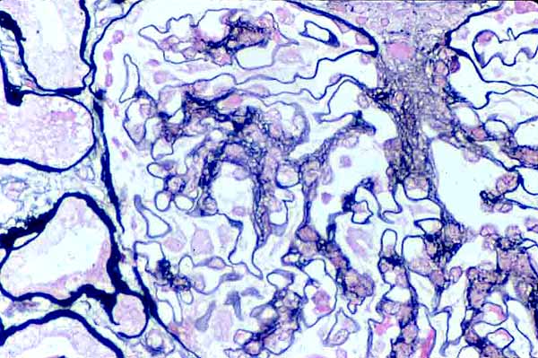
Micrograph from a needle-biopsy of renal cortex (thumbnail at right), from a patient with glomerulosclerosis. This specimen has been stained with silver to demonstrate filtration membrane and mesangial matrix.
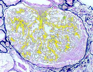
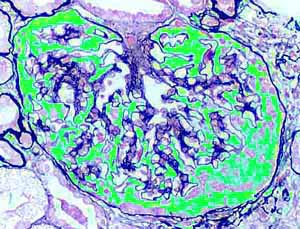
Although many features of this renal corpuscle appear reasonably normal and functional, there is a substantial increase the number of mesangial cells and mesangial matrix. (To see severely pathological glomeruli, click here or on the thumbnail at right.)
See below for interpretation of this image.
RENAL IMAGE INDEX
The vascular pole appears at the top of this corpuscle. Just above the corpuscle is a distal tubule (*) with its juxtaglomerular macula densa.
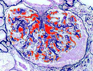
Red color highlights the lumens of glomerular capillaries. Bowman's space is uncolored. Some of the vascular space appears to be reduced by proliferation of mesangium. (Click here to see more substantially occluded glomeruli.)
Comments and questions: dgking@siu.edu
SIUC / School
of Medicine / Anatomy / David
King
https://histology.siu.edu/crr/RN084b.htm
Last updated: 16 September 2021 / dgk