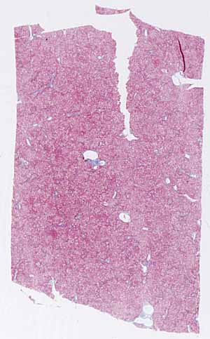
This specimen is seriously pathological. Nevertheless,
its basic architecture remains unaltered so its identity should be obvious
once you recognize the major features.
This specimen has been stained with trichrome rather than H&E.
Trichrome stains are especially useful for distinguishing collagen
fibers from other eosinophilic tissue elements. What color is
collagen in this specimen?
Note that this is a solid organ (as opposed to a hollow or tubular
one). The specimen displays cut edges all around.
Note that the entire specimen is fairly uniform, with no conspicuous
layers, regional distinctions, or subdivisions
Scattered throughout the specimen are small patches of stroma containing
a variety of tubular structures. Some of these stromal patches
show an inflammatory infiltrate (i.e., numerous small cells with round
densely-stained nuclei.)
Hints on the next page are more revealing. Don't look until
you are ready to confirm your identification.