
Fallopian Tube
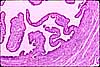
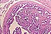
A fallopian tube (also called uterine tube or oviduct) conducts each egg, following ovulation, from the ovary to the uterus. The wall of the fallopian tube includes an elaborately folded mucosa (endosalpinx) surrounded by a muscularis (myosalpinx). The mucosa is lined by a ciliated columnar epithelium with secretory cells, and is folded out into the lumen so that an ovum in the lumen will always be close to the cilia for transport to the uterus.
- For an image of salpingitis (inflammation in the fallopian tube), see WebPath.
The size of the lumen, shape of the mucosa, and the thickness of the muscularis all vary along the length of the tube.
The mucosa is most elaborate in the ampulla, with extensive folding. The mucosa becomes much simpler, with a smaller lumen, in the isthmus. The muscularis is relatively thin (with inner circular and outer longitudinal layers) in the infundibulum and ampulla and thicker (with an additional inner longitudinal layer) as the isthmus approaches the uterus (where the muscularis merges with the myometrium.)
The mucosal lining is simple columnar epithelium, consisting of two cell types -- ciliated cells and secretory cells. Cilia, rather than muscular contraction, provide the principal motive power to move an egg toward the uterus.
Mucosal folds occupy most of the potential lumenal space, so that cilia on the epithelial surface can effectively contact the egg. Secretory cells are functionally similar to goblet cells, providing lubrication, but are not as conspicuous.
|
 |
 |
|
|
|
SIUC / School
of Medicine / Anatomy / David
King
https://histology.siu.edu/erg/oviduct.htm
Last updated: 20 May 2022 / dgk