
Areolar connective tissue, whole mount
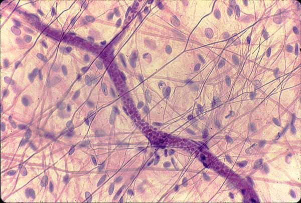
This image represents a whole mount (NOT a section) of mesentery (the tissue which binds together the loops of intestine within the peritoneal cavity).
This view looks through the entire mesentery, which consists of serous membrane (i.e., mesothelium) on front and back surfaces of this specimen, with areolar tissue and blood vessels sandwiched in between.
The two most common cell types are mesothelial cells (of the mesentery's serous membranes) and fibroblasts (of the areolar tissue).
The large, pale, oval nuclei belong to mesothelial cells on the surfaces.
The smaller, darker, more elongate nuclei belong to fibroblasts, which manufacture and secrete the molecules which form the extracellular matrix.
Macrophages, mast cells, and lymphocytes may also be found in such preparations, but none are clearly identifiable in this image.
The matrix consists of unstained ground substance through which pass fibers of made of collagen and elastin.
Elastic fibers are thin, fairly uniform in diameter, and frequently branched. The are normally visible only if specially stained (as they have been in this specimen).
Collagen fibers vary in thickness and are eosinophilic (pink, in this stain).
A conspicuous blood vessel passes across the field of view. Nuclei of vascular endothelial cells lie alongside the vessel, which contains many red blood cells.
Comments and questions: dgking@siu.edu
SIUC / School
of Medicine / Anatomy / David
King
https://histology.siu.edu/intro/IN014b.htm
Last updated: 30 December 2021 / dgk