
Lung, epithelium and connective tissue
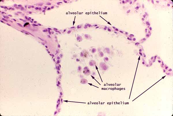
Extremely thin tissue walls separate adjacent lung alveoli. Respiratory alveoli (the large empty areas in the image) are the lung's air spaces. They comprise most of the volume of the lung.
Each alveolar wall has a simple squamous epithelium lining each exposed surface, with a thin stroma of capillaries and delicate supporting connective tissue sandwiched in between. These details cannot be clearly resolved in this image.
Monocytes from circulating blood can crawl out of the alveolar capillaries, cross the alveolar epithelium, and enter the alveolar air space. These alveolar macrophages, or dust cells, can then crawl over the free surface and scavenge dust particles and bacteria that have been inhaled. Ingested material can accumulate in lysosomal vesicles and become visible as lipofuscin granules.
Comments and questions: dgking@siu.edu
SIUC / School
of Medicine / Anatomy / David
King
https://histology.siu.edu/intro/IN015b.htm
Last updated: 1 February 2023 / dgk