
Apocrine sweat gland in skin
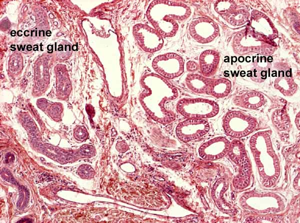

The relatively large size of the apocrine sweat gland tubule (right half of this image) contrasts with the much smaller diameter of an eccrine sweat gland on the left (duct in lower left corner). The epithelium of the apocrine gland may range from cuboidal to columnar.
To view this apocrine gland at higher magnification, click here or on the thumbnail at right.
Comments and questions: dgking@siu.edu
SIUC / School
of Medicine / Anatomy / David
King
https://histology.siu.edu/intro/apocrine1.htm
Last updated: 12 June 2022 / dgk