Skin Biopsy Specimen
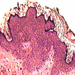
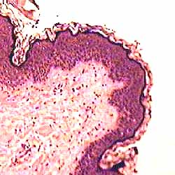
These two micrographs were taken from adjacent regions of the same specimen. (overview)
Why does the epidermis appear markedly thicker in the left-hand micrograph?
(Both micrographs are displayed at the same magnification.)
And why does a small patch of connective tissue appear to lie within the epidermis of the left-hand micrograph?
Although the epidermis appears markedly thicker in the left-hand micrograph, this does not represent an actual thickening of the epidermal tissue. This effect results from the angle of tissue orientation relative to the plane of section, which varies across the specimen.
On the left, the epidermis is cut obliquely. On the right, the epidermis is cut more-or-less perpendicular to its surface. Clear evidence for an oblique cut is provided by the small "island" of dermis which appears within the epidermis near the center of the left-hand micrograph. This is a dermal papilla which has been cut off from underlying dermis by the oblique section plane.
For more on interpreting tissue sections, see viewing tissues.
Comments and questions: dgking@siu.edu
SIUC / School
of Medicine / Anatomy / David
King
https://histology.siu.edu/intro/skinbiop/cfepi.htm
Last updated: 18 September 2021 / dgk