
Nerve cell, Golgi stain
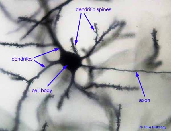
This image (© Blue Histology) shows one nerve cell in layer II of the cerebral cortex.
Dendrites are typically broad where they arise from the cell body; away from the cell body they typically taper and branch. Dendrites may extend up to a millimeter or two away from the cell body, seldom more. At the magnification shown here, the full extent of these dendrites would form a halo around your monitor. Dendritic spines represent a particular type of site for synaptic contact; they are characteristic of some but not all dendrites.
The axon (singular, one per nerve cell) is typically uniform in diameter (i.e., not tapering away from the cell body) and relatively unbranched (at least in the vicinity of the cell body). usually comparatively narrow. Axons may be many centimeters in length, up to more than a meter. This length is outrageously long, at least in terms of ordinary cellular dimensions. At the magnification shown here, the axon might extend out the door and into the next building.
For scattered individual nerve cells, the Golgi stain renders details of axon and dendrites in crisp black. But intervening nerve cells (both neurons and glia) remain unstained and invisible. This invisibility of most cells is both a strength and a weakness of Golgi stains. If all cells were similarly stained, the entire specimen would be opaque black and useless. But with only random individual cells stained, the relationships between cells (e.g., the fact that there is an axon terminal associated with each dendritic spine) are obscure.
Comments and questions: dgking@siu.edu
SIUC / School
of Medicine / Anatomy / David
King
https://histology.siu.edu/ssb/BH004b.htm
Last updated: 4 September 2021 / dgk