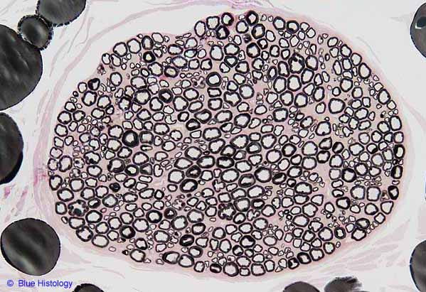

Peripheral nerve, osmium stain for myelin
Many large and small myelinated axons are visible in this cross-section of a peripheral nerve(© Blue Histology). Here each myelinated axon appears whitish (axoplasm is mostly water and generally doesn't stain well) and is sharply delineated by a surrounding black halo of myelin.
Osmium stains lipid black. Since myelin consists many layers of cell membranes, it is mostly lipid and so stains black with osmium. The several large fat droplets around the periphery of this image are also conspicuously black (rather than "empty" as they typically appear in H&E stained preparations).
Scroll down for a higher-magnification image of this nerve (also © Blue Histology). Unmyelinated axons are not apparent in either of these images. They are not only inconspicuous for lacking myelin, they are also tiny. At less than 1 µm in diameter, they are only a small fraction of the size of the smallest myelinated axon visible here. Unmyelinated axons might occur in some of the wider pale areas between myelinated axons.
Comments and questions: dgking@siu.edu
SIUC / School
of Medicine / Anatomy / David
King
https://histology.siu.edu/ssb/BH006b.htm
Last updated: 4 September 2021 / dgk