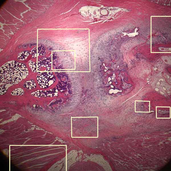

Fibrocartilage and bone (pubic symphysis)

At the center of this image is fibrocartilage of the pubic symphysis. On either side is bone, with endochondral ossification occurring at the boundaries.
- Bone is deep red.
- Cartilage is variously gray with pink (collagenous) bands.
- The spongy tissue (dark purple with white spots) is bone marrow with fat cells.
- In the four corners is skeletal muscle attached to the periosteum / perichondrium.
Click on one of the frames above, or one one of the thumbnails below, for detail. Or choose the thumbnail at right for a labelled view.
Comments and questions: dgking@siu.edu
SIUC / School
of Medicine / Anatomy / David
King
https://histology.siu.edu/ssb/NM003b.htm
Last updated: 12 August 2021 / dgk