
Motor Neuron, Spinal Cord Smear
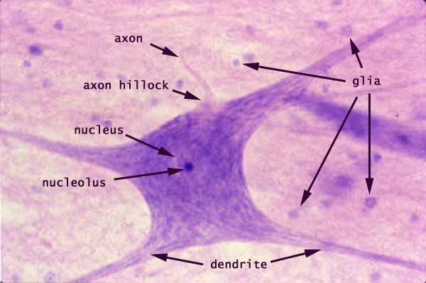
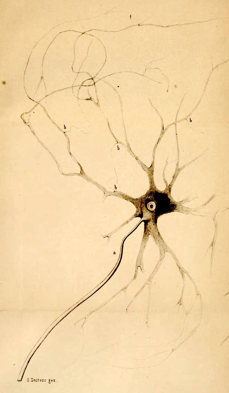
This slide image represents a smear preparation of spinal cord grey matter, in which a small bit of nervous tissue has been squished onto the slide (rather than neatly sectioned). Once a cell body has been found, its (flattened) dendrites can be seen extending in several directions. (This micrograph might be compared with an impressive artistic rendering from the nineteenth century, at right.)The conspicously large cell is a motor neuron from the anterior horn. Several features may be noticed.
The cytoplasm is rather coarsely textured, with the darker (basophilic) patches representing rough endoplasmic reticulum ("Nissl bodies").
The large nucleus is obscured by overlying cytoplasm, but the densely-stained nucleolus remains distinctly visible.
A thin axon arises from an axon hillock. Both are recognizable by their pale axoplasm, which is pale because basophilic ribosomes are absent from axoplasm.
[In spinal smears, axons are only noticeable when they happen to fall along the edge of the flattened cell bodies. Most axons will be squished against the tops or bottoms of their cells, and will thereby be practically invisible.]
Dendrites extend away from the cell body beyond the edges of the image. Cytoplasm within the dendrites is similar to that in the cell body (i.e., containing dark Nissl bodies, in contrast with the pale cytoplasm in the axon).
The background texture comprises a tangled feltwork ("neuropil") of dendrites and axons from many nerve cells, as well as glial cells and their processes. The numerous small nuclei throughout the field of view belong to glial cells
Comments and questions: dgking@siu.edu
SIUC / School
of Medicine / Anatomy / David
King
https://histology.siu.edu/ssb/motoneur.htm
Last updated: 12 June 2025 / dgk