 |
Please watch patiently to see the animation. from |

Please watch patiently to see the animation.
Osteoclasts are pink.
Osteoblasts are blue.
Bone lamellae are gray.
Osteocytes (within the lamellae) are not shown.from
Blue Histology,
Copyright Lutz Slomianka 1998-2004
Notes
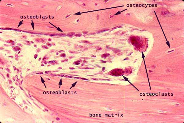
These images illustrate the process of bone remodelling.
Note the sequence of events. Compare the micrograph, with remodelling proceeding toward the right, with the cartoon remodelling that proceeds toward the left
Osteoclasts remove bone matrix, enlarging a channel through the bone. (The apparent separation between the osteoclasts and the adjacent bone matrix is an artifact of preparation.)
Osteoblasts lie alongside the surface of the channel, where they are laying down new lamellae. They follow after the osteoclasts, rebuilding the bone (and thereby storing calcium). Eventually most of the space, except for a narrow Haversian canal, will be occupied with fresh bone.
A balance between osteoclast and osteoblast activity is necessary for a stable calcium level in blood. To this end, osteoclast activity is hormonally regulated, stimulated by parathyroid hormone and inhibited by calcitonin from C-cells of the thyroid.
Osteocytes are embedded in the bone matrix, each within its own lacuna. As remodelling proceeds, osteocytes are alternatively freed and then re-imprisoned.
Lamellae lie parallel to freshly-formed surfaces (i.e., immediately beneath the osteoblasts), but are cut arbitrarily by osteoclast activity.
Comments and questions: dgking@siu.edu
SIUC / School
of Medicine / Anatomy / David
King
https://histology.siu.edu/ssb/remodel.htm
Last updated: 9 August 2021 / dgk