
Trachea, epithelium and connective tissue
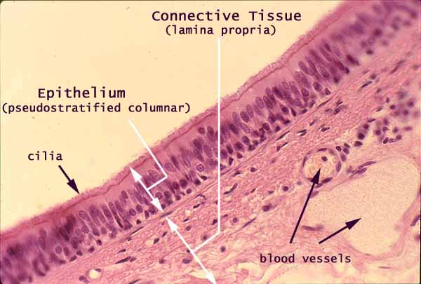
This image shows the pseudostratified columnar epithelium that lines the mucosa of the trachea. This epithelium consists of both short and tall cells, all resting on the basement membrane.
- Most of the tall (columnar) cells are ciliated, a specialization that allows dust and mucous to be swept upward and eliminated.
- Mucus-secreting goblet cells are not apparent in this image.
The tracheal epithelium is supported by loose connective tissue.
The loose connective tissue of a mucosa is called lamina propria, and typically has a fairly high proportion of cells with immune function.
Within the lamina propria, the small round nuclei may belong to lymphocytes. The more elongated nuclei are most likely fibroblasts.
Comments and questions: dgking@siu.edu
SIUC / School
of Medicine / Anatomy / David
King
https://histology.siu.edu/crr/CR006b.htm
Last updated: 15 June 2022 / dgk