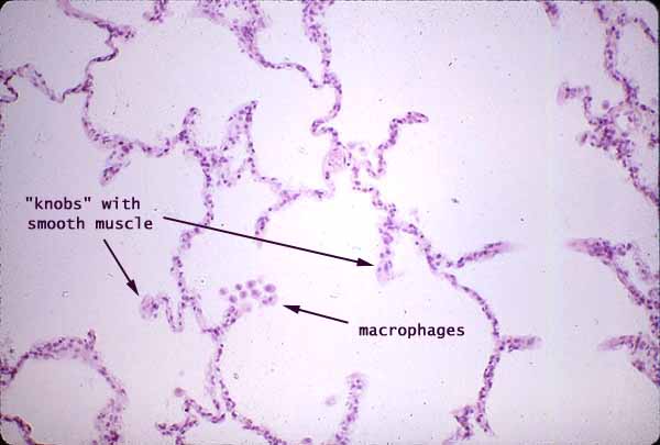

Lung alveoli

Most of the volume of the lung consists of air-filled space, subdivided into millions of small interconnected chambers by alveolar walls. The cellular composition of alveolar walls cannot be readily resolved at this magnification. (In this specimen, the shape of the alveoli is somewhat distorted by preparation.)
At the entrance to each alveolus, a "knob" along the alveolar wall contains smooth muscle fibers which allow the size of the opening to be adjusted. Each alveolar wall is lined on either surface by simple squamous epithelium, with capillaries sandwiched in between.
A small cluster of alveolar macrophages ("dust cells") may be seen near the center of the image
Comments and questions: dgking@siu.edu
SIUC / School
of Medicine / Anatomy / David
King
https://histology.siu.edu/crr/CR017b.htm
Last updated: 27 May 2022 / dgk