Renal corpuscle, electron micrograph
 |
| Micrograph courtesy of Jeff Morton |
See below for a labelled image.
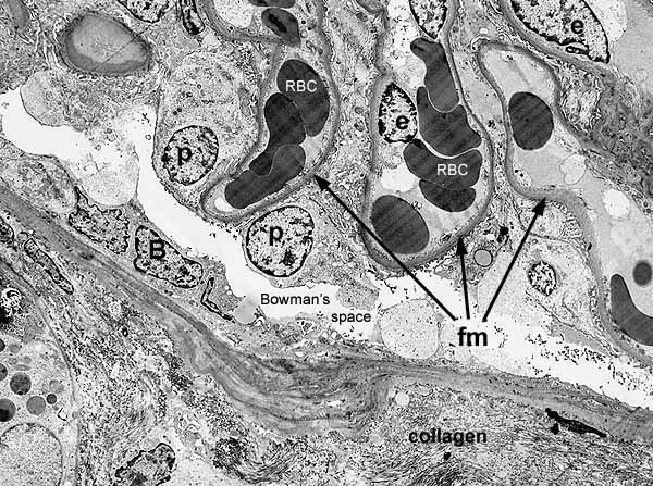
The cortex of the kidney is distinguished by characteristic renal corpuscles, each of which consists of an outer envelope (Bowman's capsule) surrounding a fluid-filled space (Bowman's space) within which is suspended a glomerulus.
This micrograph includes portions of several glomerular capillaries and, toward the bottom of the image, Bowman's capsule with underlying cortical stroma. For greater detail, click on the thumbnail at right.
RBC = red blood cell in capillary lumen
fm = filtration membrane
p = nucleus of podocyte
e = nucleus of capillary endothelium
B = nucleus of Bowman's capsule epithelium
Collagen appears in the interstitial space below the basement membrane of Bowman's capsule
Micrograph courtesy of Jeff Morton
Comments and questions: dgking@siu.edu
SIUC / School
of Medicine / Anatomy / David
King
https://histology.siu.edu/crr/EM002b.htm
Last updated: 13 September 2021 / dgk