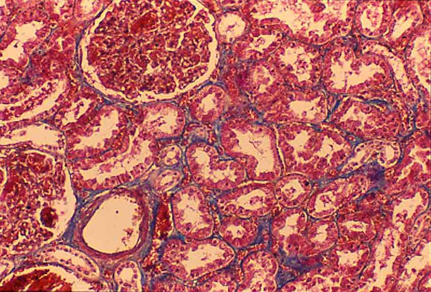


Trichrome stains make renal stroma more apparent, by selectively staining collagen (blue in this specimen).
(Renal parenchyma consists almost entirely of tubule epithelium and comprises the bulk of the kidney. In H&E-stained specimens [most of the micrographs in this website], supporting stromal tissue (collagen and capillaries) is inconspicuous.)
Trichrome staining is especially useful in pathology by clearly revealing the amount of collagen. An increase in collagen could indicate a history of scarring after tissue loss.
PAS
stain
Comments and questions: dgking@siu.edu
SIUC / School
of Medicine / Anatomy / David
King
https://histology.siu.edu/crr/RN029b.htm
Last updated: 15 September 2021 / dgk