
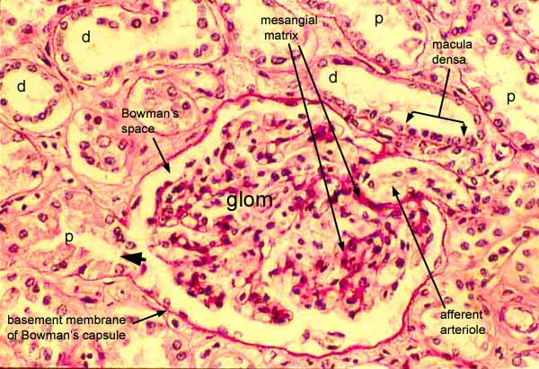
The cortex of the kidney is distinguished by characteristic renal corpuscles, each of which consists of an outer envelope (Bowman's capsule) surrounding a fluid-filled space (Bowman's space) within which is suspended a glomerulus (glom).
Bowman's space may be seen opening into the beginning of a proximal tubule (arrowhead) at the urinary pole of this corpuscle.
The macula densa may be seen at the vascular pole of the corpuscle, displaying its characteristic appearance of several distal tubule nuclei crowded densely together. Nearby is the associated afferent arteriole.
The PAS stain accentuates the filtration membrane as well as the basement membrane of renal tubules and Bowman's capsule and also the mesangial matrix.
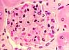
Although the glomerulus in this image clearly contains many cells with varied appearances, their individual identities as endothelial cells, podocytes, or mesangial cells are difficult to determine reliably on relatively thick-sectioned specimens such as this.
The lumens of distal tubules (d) commonly appear more open and clear than those of proximal tubules (p).
Note that fixation of this specimen is not ideal, such that some epithelial cells have become detached from their underlying basement membrane.
Comments and questions: dgking@siu.edu
SIUC / School
of Medicine / Anatomy / David
King
https://histology.siu.edu/crr/RN046b.htm
Last updated: 29 May 2022 / dgk