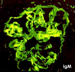
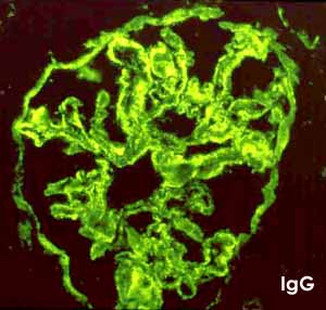
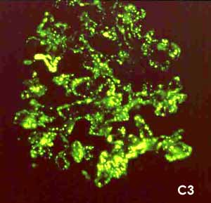
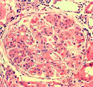
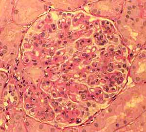
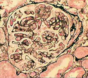
Renal glomeruli, membranoproliferative glomerulonephritis
These images portray renal glomeruli as seen with six different stains, all of which reveal to a greater or lesser extent the accumulation of material in the area of the mesangium and glomerular basement membrane, associated with membranoproliferative glomerulonephritis. (All six images are from the same clinical case.)
For related images with additional interpretation of glomerular organization, click on the thumbnail at right.
For additional discussion and images of renal disease, see WebPath. (This page is worth an extended visit, after you have become comfortable with basic kidney histology.)
Comments and questions: dgking@siu.edu
SIUC / School
of Medicine / Anatomy / David
King
https://histology.siu.edu/crr/RN124b.htm
Last updated: 16 September 2021 / dgk